
Questions? Or do you want me to blog on a specific topic? Email me at: drchris@dpt.services
Want to get the latest blog posts? Sign up for the Thursday Three Newsletter below.
What Exactly is an Electrolyte?
An electrolyte is a mineral that carries a charge when dissolved is a fluid such as water or blood. When you ingest food or beverage, it is broken down into smaller parts. Some are broken into neutral atoms or molecules, and some are broken into charged atoms or molecules. If they have a charge, they are called ions, or electrolytes.

An electrolyte is a mineral that carries a charge when dissolved is a fluid such as water or blood. When you ingest food or beverage, it is broken down into smaller parts. Some are broken into neutral atoms or molecules, and some are broken into charged atoms or molecules. If they have a charge, they are called ions, or electrolytes. The electrolytes of the body are:
Sodium (Na+)
Potassium (K+)
Calcium (Ca2+)
Bicarbonate (HCO3-)
Magnesium (Mg2+)
Chloride (Cl-)
Phosphate (PO4 3-)
The human body is a bioelectric machine, and is capable of producing energy equivalent to a 100 watts light bulb. Electric currents in the body are used by different functions in the body such as conducting nerve impulses, controlling metabolism, and regulating tissue and organ function.
Electrolyte balance is crucial for normal human functioning and helps maintain fluid levels in cells. If the electrolyte concentration in a cell is too high, fluid will move into that cell via osmosis. If the electrolyte concentration is too low, fluid moves out of the cell. This is why your pee is very yellow when you are dehydrated; the body is trying to maintain electrolyte balance and dumps electrolytes to restore balance.
The kidneys are the main organ that filters electrolytes and maintains balance. If this system is disrupted, dysfunctions can occur. Let’s look at salt, since it’s the main electrolyte. Too much salt (hypernatremia) can cause thirst, confusion, irritability, fatigue, spasms or muscle twitching. Too little (hyponatremia) can cause dizziness, headache, spasms, and even seizures or coma. Sounds serious, but the good news is that it’s relatively easy to maintain balance as long as your kidneys are functioning well. You can use a urine color chart to monitor your hydration status:

But wait, I thought alcohol dehydrates you, so why does my pee turn clear when I have alcohol? It is because the kidneys are guided by hormones secreted from the pituitary gland in your brain. Alcohol is able to cross the blood brain barrier and disrupt the hormone signaling. ADH is a hormone that tells the kidneys to hold on to water, and alcohol inhibits this hormone. So the whole function of the kidney is altered and the body starts wrongly dumping water. Then in the morning, when the alcohol is no longer affecting your brain, the kidneys start working normal again and recognize a gross electrolyte imbalance and dump salts, making your pee very yellow. Now you are depleted in water and electrolytes, so you become very thirsty and crave salty food. This is how brunch was born. Drinking water with every alcoholic beverage can help buffer this to an extent.
References:
Fluid and Electrolyte Balance. (2020, October 01). Retrieved October 17, 2020, from https://medlineplus.gov/fluidandelectrolytebalance.html
Nguyen, T. (2014, March 24). This Flashlight Is Powered by the Touch of Your Hand. Retrieved October 17, 2020, from https://www.smithsonianmag.com/innovation/this-flashlight-is-powered-by-the-touch-of-your-hand-180950226/
User, S. (n.d.). The Science of Bioelectricity. Retrieved October 17, 2020, from https://www.e-qure.com/the-science-of-bioelectricity/the-science-of-bioelectricity

CO2 Tolerance
Carbon dioxide (CO2) has a bad rep. Classically we are taught that when you breathe, blood cells become oxygenated in the lungs, and then the oxygen is delivered to the organs and tissues, and carbon dioxide is then exhaled as a waste product. Although that is not wrong, it’s missing some key steps. So more oxygen (O2) is what we need right? Take a big deep breath, right? Well the issue is not the amount of oxygen in the blood, but delivering the oxygen to the tissues so it can be used. Turns out, carbon dioxide is a big factor, and our tolerance to carbon dioxide is crucial to athletic performance.

Carbon dioxide (CO2) has a bad rep. Classically we are taught that when you breathe, blood cells become oxygenated in the lungs, and then the oxygen is delivered to the organs and tissues, and carbon dioxide is then exhaled as a waste product. Although that is not wrong, it’s missing some key steps. So more oxygen (O2) is what we need right? Take a big deep breath, right? Well the issue is not the amount of oxygen in the blood, but delivering the oxygen to the tissues so it can be used. Turns out, carbon dioxide is a big factor, and our tolerance to carbon dioxide is crucial to athletic performance.
Oxygen levels (or saturation) in the blood doesn’t change much unless you have a breathing dysfunction like COPD. It might bounce between 95% and 98%, not a drastic change. So why do we feel the need to take deeper breaths during exercise? It may be due to a sensitivity to accumulation of CO2.
A quick lesson: Blood cells transport both O2 and CO2. When a blood cell is in an area where there is higher concentration of one of these molecules, it will pick up more of said molecule. In the lungs there is higher O2, so the blood picks up O2. In the muscle tissue, there is more CO2, so it picks up more CO2, and the O2 is offloaded to the muscles. As this happens, the CO2 concentration rises and there are receptors in the vessels that monitor this. If they are sensitive, they will stimulate the urge to breathe sooner and less O2 is offloaded to the muscle. So the key is to build your tolerance to CO2 accumulation so more O2 can be offloaded and used. This can have a dramatic effect on athletic performance.

There is a simple test you can perform to assess where you stand with CO2 tolerance; it’s called the BOLT test. Here is how it works:
Take a normal breath in through your nose and a normal exhale out through your nose.
Hold your nose to prevent air from entering your lungs.
Count the number of seconds until you feel the first definite desire to breathe, or the first stresses of your body urging you to breathe. This may include a need to swallow, or a constriction of the airways, abdomen, or throat.
Release your nose and breathe in through your nose. You should be able to resume normal, calm nasal breathing immediately. If you need to take a big mouth breath, you have held your breath for too long.
Average tends to be around 20 seconds. 25-40 seconds is ideal. If you are under that, you may be sensitive to CO2. The good news is you have room to grow. Lower scores are associated with anxiety and stress. This is a good litmus test and can be a snapshot of where you are currently. If work has been stressful, you can expect a lower score.
So how do we improve CO2 tolerance? Free divers have been exploring this for years and there is a dearth of knowledge out there with different techniques. But there are a few simple techniques. Nasal breathing is key, and you can perform light aerobic activity like walking with nose only breathing. Nasal breathing will increase nitrous oxide in your body which is a strong vasodilator. Nasal breathing also activates the diaphragm and slows down breathing. Faster breathing tends to lead to chest breathing.
You should also have a daily breathing routine. Take about 10 minutes a day and you can either work on cadence breathing, which means only 6 breaths per minute with longer exhales. Or you can put your hands on your chest and stomach, slow down the speed of air coming in your nose and out of your nose. When you feel the need to breath, take a slow breath in. This is a great exercise in the evening prior to bed.
You may feel warm after doing this. That is because CO2 is a vasodilator and allows oxygen to pass into the tissues easier. When we get short of breath and begin breathing too hard and get rid of too much CO2, the blood vessels constrict, and the bond between hemoglobin and oxygen is strengthened.
For more information, check out Patrick McKeown. He wrote, “The Oxygen Advantage”. If you aren’t a reader, you can check out this podcast:
SCP Podcast 168: Oxygen Advantage with Patrick Mckeown

Coughing and Back Pain
I recently had bronchitis and had several days of intense coughing. Shortly afterwards, I developed upper and lower back pain. I even started getting mild numbness in the pinky finger on that same side. It happened to be my left side. The pain extended up into my neck as well and felt like a deep ache. Tell a healthcare provider this set of symptoms, and they are instantly going to be worried about a heart attack. Lucky for me, it was just an irritated muscle.

I recently had bronchitis and had several days of intense coughing. Shortly afterwards, I developed upper and lower back pain. I even started getting mild numbness in the pinky finger on that same side. It happened to be my left side. The pain extended up into my neck as well and felt like a deep ache. Tell a healthcare provider this set of symptoms, and they are instantly going to be worried about a heart attack. Lucky for me, it was just an irritated muscle.
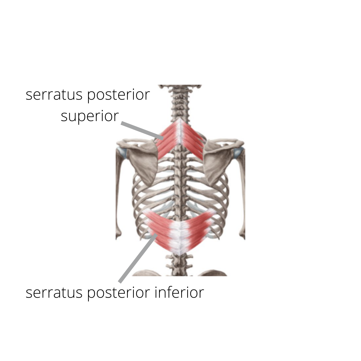
The serratus posterior is a deep muscle, and lies underneath the big upper traps. It connects the vertebrae (C7-T2 or T3) to the ribs 2-5. It’s job is to elevate the ribs so it’s active in respiration. With all of my coughing fits, those muscles were working hard and went into overuse. I also tend to be stiff in my thoracic spine, so with the muscle rapidly contracting, coupled with the fact that one end of the muscle is tethered down to a stiff vertebrae, it quickly got irritated. I also developed pain in the lower serratus posterior inferior muscle, but I’m not stiff in the lumbar spine so this pain went away on its own.
So how did I solve this? Mostly by working on mobilizing the irritated muscle, my thoracic mobility, and with breathing. Let’s break it down:
I first wanted to desensitize the irritated tissue because I could barely turn my head or lift my arm overhead, so I layed on a foam roller in the direction of the muscle fiber and worked on those two movements. I turned my head slowly from side to side. Each time I got to my end range, I made sure to take several deep breaths. Each repetition, I aimed for more range of motion. Then I did the same process but this time I reached overhead with my arm. It took about 5 minutes.
My upper back tends to be stiff in extension and rotation. So I did these two drills:
Then to finish everything off, I just layed down on my back and worked on some breathing drills. I focused on a long exhale as this is the eccentric phase of the muscle; so in other words, it elongated it. Within 10 minutes or so, I was pain free and my range of motion was restored.
This is a perfect example of how precision can make all the difference in a rapid recovery.

Breathe
One of the simplest exercises someone can do when in pain is breathe. It sounds a bit silly, but you’d be surprised at how effective it is. Stay with me. First, let’s have a brief discussion about pain. Pain is a warning signal from the brain to change something. It can be acute or chronic, it doesn’t matter; the nervous system is sensing something is a threat. In other words, your flight or fight mechanism is turned on (the sympathetic nervous system).

One of the simplest exercises someone can do when in pain is breathe. It sounds a bit silly, but you’d be surprised at how effective it is. Stay with me.
First, let’s have a brief discussion about pain. Pain is a warning signal from the brain to change something. It can be acute or chronic, it doesn’t matter; the nervous system is sensing something is a threat. In other words, your flight or fight mechanism is turned on (the sympathetic nervous system).
If you take a few minutes to lay down and concentrate on your breathing, it can stimulate the relaxation response (parasympathetic nervous system). The alarm bells are turned off. If you can lay down in a manner that is pain free, it’s even better. This alone is a powerful tool, but let’s go over some other reasons breathing can reduce pain or tension.
Breathing helps reperfusion. Through our daily life, we develop waste products. This could just be the stress of life, or working out, there is always waste product and it’s normal. This is flushed out through our lymphatic system. Blood brings new oxygen to muscle tissue and this process occurs through the cardiopulmonary system. When there is a painful area in the body, there is often congestion in the area, i.e. waste product build up that needs to be decongested. Have an ache in the back? Spend a few minutes deep breathing and it may reduce that pain simply by decongestion and reperfusing oxygenated blood to the area.
You also have gentle, non-threatening movement. When you inhale, your hips anteriorly tilt and your spine extends. When you exhale, your hips posteriorly tilt and your spine flexes. Reteaching the brain that the spine and hips can move without pain can do wonders for pain reduction, especially in situations such as chronic pain or when the body has been sensitized.
Gas exchange is also a benefit. So many of us through our stressful work/life environments, develop a habit of shallow breathing. When you don’t fully exhale, you leave used up air in the lungs in the form of carbon dioxide. When you don’t fully inhale, you don’t get to take advantage of all that good oxygen.
There are conditions like COPD and asthma that lead to dysfunctional breathing patterns. In some cases, the diaphragm becomes inactive and the person develops chest only breathing. Then all the muscles that attach to the ribs like the pecs and neck muscles have to do the work by actively pulling the ribs up. This can lead to all kinds of fun neck and shoulder pain. The belly and diaphragm are supposed to move when you breathe. Ever notice a woman’s neck muscles when they are wearing a tight dress or corset? They have temporarily induced chest only breathing by restricting the belly.
So how do you do it correctly? Get comfortable on your back, take a deep breath in through your nose, then exhale through your mouth 3 times as long as you inhale. As you inhale, your belly should rise, then your ribs should expand both on your sides and at your chest. Think of the abdominal cavity as a canister that expands and contracts. Try it for at least 2 minutes. Notice areas of your body that may feel stiff and see if anything changes.
This is a great way to wind down the day. As a society, we are great at stepping on the gas pedal. Rushing to work, energy drinks, Starbucks everywhere, work, then go right to the gym, etc. What we are not great at doing, is turning on the brakes and slowing down. Put deep breathing into your bedtime habit, and even the fact that you are disengaging from technology, and spending a few minutes on yourself can be a game changer. Who knows, you may sleep better.

Why I Don’t Stretch Before Hockey
I’ve had a number of teammates ask me why I don’t stretch before I skate. It’s a relatively complicated answer, but the main reason is to avoid injury. Let me explain:

I’ve had a number of teammates ask me why I don’t stretch before I skate. It’s a relatively complicated answer, but the main reason is to avoid injury. Let me explain:
First let me clarify that I don’t do passive stretching prior to a game. This is the classic leg on the board, stretch the hamstring, or knees on the ice groin stretch. I do a form of active stretching which I’ll get into later.
You have to ask yourself, why am I stretching? The classic wisdom was to avoid injury, but there is no evidence that this is true. There is however, evidence that it decreases athletic performance. One study found that stretching the hamstrings prior to a one rep max hamstring curl reduced performance (Winchester et al., 2009). Now these effects are probably transient, so I don’t put too much stock in that study, but it just adds to my reasoning of why I don’t stretch before I play.
Passive stretching is effective in creating a temporary increase in range of motion of a joint. There is something that is called the stretch reflex, and it’s protective. It arises from the nervous system and is meant to prevent muscles from tearing. Picture yourself carrying a heavy box, then your buddy throws a 20 lb dumbbell into the box. Your biceps would respond by quickly activating. That is the stretch reflex. When the reflex is exposed to repeated stimulus, i.e. passive stretching, the excitability of the reflex decreases and you have a sudden, albeit temporary, increase in range of motion (Guissard & Duchateau, 2006). The problem with this is the new range of motion is not stable; you don’t have good control or strength at this new end range. This is where injuries occur. Picture a soccer player who has to rapidly extend the knee to kick a ball. The hamstring has a role in limiting this movement. If the soccer player spent some time stretching the hamstring before the game, the knee will now overextend and this is a great way to rupture an ACL.
I do think it is important to improve mobility. Notice I didn’t say flexibility. Mobility is flexibility plus the ability to be strong in the end range. So that means training strength after gaining new range of motion. This should be a work in progress, and should happen in a controlled environment. Sports are the opposite of a controlled environment. It’s not like you are thinking that you should be working on your form during a game, that’s what practice is for. I do stretch my calves before I squat, but that is a controlled environment; I’m concentrating on my form.
So what to do before a game? Active stretching. What is the difference? The goal of active stretching is to bring your joints to your normal end range, not past it. So it’s self limiting. It also gets blood flow to the muscles and gets them warm and ready for action because it uses muscle contraction. What does this look like? I’ll use my stick and rotate my wrist to warm up the forearms for stickhandling. I’ll twist my torso to get my core and back ready for taking shots. I’ll carve C’s in the ice with my legs to get ready for skating. And I’ll skate, that’s technically an active warm up too.
When you’re young, you can get away with passive stretching but us older beer league hockey players should be a bit more mindful of how we prepare ourselves.
References:
Winchester JB, Nelson AG, Kokkonen J. A single 30-s stretch is sufficient to inhibit maximal voluntary strength. Res Q Exerc Sport, 2009; 80: 257-261.
Guissard, N., & Duchateau, J. (2006). Neural Aspects of Muscle Stretching. Exercise and Sport Sciences Reviews, 34(4), 154-158. doi:10.1249/01.jes.0000240023.30373.eb

3 Reasons Your Joints Pop
It’s a common complaint I hear; my knees pop when I squat or my hip snaps. People often seem concerned about this, but generally speaking, this is considered normal.There are 3 typical reasons joints make sound: 1. Tendons, 2. Arthritis, 3. Gas bubbles.

It’s a common complaint I hear; my knees pop when I squat or my hip snaps. People often seem concerned about this, but generally speaking, this is considered normal.There are 3 typical reasons joints make sound: 1. Tendons, 2. Arthritis, 3. Gas bubbles.
Tendons are the ends of the muscle and serve as attachments to bone. Because of this, they are located near bony prominences. Sometimes they roll over these areas like in snapping hip syndrome. Other times, they roll over other tendons such as the rotator cuff which is a series of 4 tendons. The general rule of thumb is if it doesn’t hurt, it’s nothing to worry about. However, if you are doing shoulder raises and your shoulder is popping with every rep, change the rotation of your shoulder (by changing your hand position) and see if you can get it to go away.

Another cause of sound is arthritis. At the ends of our bones, we have cartilage which is a slippery substance and allows joints to glide smoothly on each other. As we age, the cartilage thins or wears away and now the jagged edge of the bone grinds on the neighboring bone. This is very common in the spine, particularly the neck.
The last reason, and probably the most common, is the good ol’ gas bubbles. This is what you hear when you crack your knuckles, or when a chiropractor or physical therapist manipulates your spine. There is controversy surrounding this topic. The prevailing theory for years was that a bubble of gases inside the joint pops or collapses, causing the sound. This is called cavitation and is said to give you a sudden increase in mobility. A study in 2015 by Kawchuk et al. found just the opposite. Through real time MRI, they were able to conclude that the sound is actually the formation of a bubble when the joint is stretched, rather than a collapse. There is a substance called synovial fluid that is inside the joint and has an adhesive property. When force is applied to the joint that is stronger than the adhesion, the fluid fractures and causes a cracking sound. This is called tribonucleation. The idea here is that when the joint separates, negative pressure is formed and pulls dissolved gases out which form a bubble.

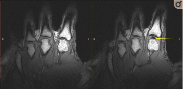
Kawchuk et al., 2015
That begs the question, is it good or bad for you? Well as we understand it now, it’s neither good or bad for you. There is no evidence to suggest that it causes arthritis. As a physical therapist, we manipulate joints to restore range of motion, the sound is just a by-product and is not an indication of whether the mobilization was successful or not. We will usually tell you to not attempt this on your own. The way the body works is that if you are stiff in one area, you tend to be “loose” or hypermobile in the neighboring area. If you like to crack your own neck, you are probably making a loose area looser. When we do it, we are doing our best to be specific to the stiff area. Although this is not an exact science, it’s certainly better when targeted.
References:
Kawchuk, G. N., Fryer, J., Jaremko, J. L., Zeng, H., Rowe, L., & Thompson, R. (2015). Real-Time Visualization of Joint Cavitation. PLOS ONE, 10(4), e0119470. https://doi.org/10.1371/journal.pone.0119470
Questions? Or do you want me to blog on a specific topic? Email me at: drchris@dpt.services

Are You Metabolically Inflexible?
Your body can produce energy from multiple sources, the two most common being carbs and fats. If you are able to easily switch between the two sources without crashing, you are metabolically flexible. If, for years, you’ve been on a high carbohydrate diet, chances are you are not metabolically flexible.

Did I get your attention?
Metabolic flexibility is a term that’s being thrown around a lot right now, and for good reason. With the current COVID pandemic, there appears to be a strong predilection towards impacting people with metabolic syndrome which is directly correlated to metabolic inflexibility. Metabolic syndrome is a cluster of symptoms which appear to tie back to insulin resistance, a.k.a. Diabetes. Millions of Americans are diabetic, and millions more are prediabetic.
What do these terms mean? Your body can produce energy from multiple sources, the two most common being carbs and fats. If you are able to easily switch between the two sources without crashing, you are metabolically flexible. If, for years, you’ve been on a high carbohydrate diet, chances are you are not metabolically flexible. The problem is, following Eisehower’s heart attack in 1955, a physician named Ancel Keys convinced the American Heart Association that a diet high in saturated fats leads to heart disease. Shortly after, a diet of low fat and high carbs was recommended. Years later, the United States skyrocketed in incidences of diabetes, stroke, and childhood obesity; and the numbers continue to climb.
I remember in the 90’s, hearing that I could lose weight by eating as much meat and cheese as I wanted, but no rice or pasta. I couldn’t believe it. The Atkins diet had surfaced. Over the years, this low carb, high fat diet has morphed into the Paleo diet, and now the Ketogenic diet. If you think about it, all this is, is a return to the way humans have evolved to eat over millennia. The Inuit (Eskimos), have traditionally lived off of a diet of primarily whale blubber and have some of the lowest incidence of chronic diseases on the planet.
So what is going on here? Why are carbs being taken off the table? The bigger part of this is that Americans have been eating an overabundance of carbs. And I mean refined carbs. The carbs from fruit and vegetables are not evil, and they carry the additional benefit of being loaded with micronutrients. It’s the white bread, rice, pasta, etc. The problem with using simple carbs as a fuel source, they can glycate (stick to) our blood cells. This can cause oxidation.
Oxidate stress can wreak havoc, eating away at the lining of our blood vessels, leading to clots and strokes. By the way, the oxidation process of free radicals is neutralized by antioxidants. In other words, fruit and vegetables. See how it all ties together? A quick tip, the darker the fruit, the more antioxidants. If a fruit browns, it is low in antioxidants. The process of oxidation leads to browning (think of apples and bananas left out). Just like when iron is exposed to oxygen, it rusts, I think of oxidation in humans as our way of rusting.

The constant flux of carbs in the system leads to insulin resistance. Insulin is a hormone that transports carbs into cells. If the body chronically gets excess carbs, the insulin can’t keep it’s job up. The brain can become insulin resistant as well and this is called type 3 diabetes. This leads to damage to the nerves and it’s thought that a carb heavy diet can lead to Alzheimer’s.
This brings me to the next trend, fasting. The idea here is that once the body runs out of carbs to burn, it turns to fat stores. Fat breaks down into ketone bodies for fuel and now you are in a state of ketosis. This is where Ketogenic diet gets its name. An easy way to tell if you are metabolically inflexible is if you get ‘hangry’ when fasting.
So the bottom line is that we want to become metabolically flexible. You are just a more adaptable being if you can use all food sources for energy. Also, if you get energy from a more normal level of carbs, you don’t run the risk of insulin resistance and the cascading damage of free radicals.
How does one do this? Choose whole foods and don’t eat things that are white or that don’t rot. If it’s got a list of ingredients, it’s probably bad. Try to achieve a state of ketosis, so maybe the ketogenic diet or intermittent fasting is for you. Also, surprise surprise...exercise. Exercise has been demonstrated to improve insulin sensitivity.
Want to get a hold of your metabolic health? That’s what we seek do with functional medicine. Hop on a free call to learn more:
Running Series: Neck and Shoulder Pain
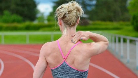
This is an area of the body that is not often associated with pain when running, but for those that experience it, it can be a confusing nuisance. Usually the underlying problem stems from stiffness in the thoracic spine, or mid to upper back. This most often occurs with anyone who sits at a desk for work.
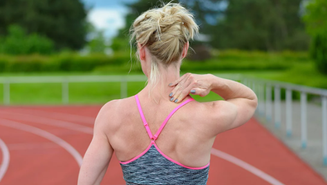
This is an area of the body that is not often associated with pain when running, but for those that experience it, it can be a confusing nuisance. Usually the underlying problem stems from stiffness in the thoracic spine, or mid to upper back. This most often occurs with anyone who sits at a desk for work. The typical hunched posture at work tends to lead to a lack of extension and rotation through this part of the spine. It is, by the way, the large part of the spine and plays a role in transmitting force from the legs to the torso or vice versa.
When I watch someone running and see that their shoulders are hunched up, or their arms swing almost side to side rather than front to back, it’s a telltale sign of a stiff thoracic spine. Rotation is important in running for balance and momentum. As one leg strides forward, the opposite side arm swings backward and the thoracic spine rotates. If there is restriction in rotation, funny things happen to arms such as a popeye style swing to their arms due to lack of rotation in their spine.

Lack of extension (think leaning back) will cause issues as well. A rounded upper back will cause a hunched up appearance to the shoulders as the shoulder blades are not able to slide down on the rib cage. This will tend to lead to pain in the upper traps and particularly the levator scapula.

How do we fix this besides quitting your job? For thoracic extension, I recommend buying a foam roller, which is just a great tool to have anyway.
For rotation, here is a simple exercise to mobilize the spine:
If you want to learn more, we have launched a running program. Please submit your info here to get on the waiting list.
Running and Back Pain
One of the more frequent complaints I hear from runners is back pain after a run. There may be many sources of this pain, but the first thing I’ll assess are the hip flexors.
One of the more frequent complaints I hear from runners is back pain after a run. There may be many sources of this pain, but the first thing I’ll assess are the hip flexors.
Tight, or shortened hip flexors can cause issues in both running and walking. Whatever a muscle does (hip flexion in this case), limits the opposite motion (hip extension). This is hip extension:

Both walking and more so running, require a fair amount of hip extension. So if the hip flexors are not allowing for adequate range of motion, the body will find it somewhere. The hip flexors are a group of muscles that originate on the sides of the vertebrae so as the leg extends back, and the hip flexors run out of laxity, it will pull forward on your vertebrae. This can be even worse if only one side is tight, because now it will also put a rotational force on the vertebrae.

The iliopsoas muscles are the hip flexors. Note the attachment to the side of the vertebrae
There is another muscle, rectus femoris, that is considered a weak hip flexor. This muscle starts from the top of your pelvis and attaches below your knee cap; it’s a two joint muscle. This one can also be problematic if one side is more restricted than the other and can lead to SI dysfunction (sacroiliac) which will also mimic back pain.
Usually a combination of manual techniques similar to massage, and lengthening these muscles will resolve the issue. It will also be necessary to strengthen the core to prevent too much movement of the vertebrae or SI joint. Also, it will be important to learn how to extend the hip without creating too much of an arch in your lower back.
Questions? Or do you want me to blog on a specific topic? Email me at: drchris@dpt.services
You Are Not Your MRI; Myths About Low Back Pain
When you dig for dirt, you find dirt. This is how it is with imaging. Stenosis, spondylosis, degenerative disc disease, degenerative joint disease, schmorl’s nodes, foraminal narrowing, osteophyte formation; these are all scary words. Here’s the thing, they’re all normal. They are just words for aging.
This is all too common of a story; a patient has severe back pain. The pain is so bad that the patient has to lay in bed for a day or two and take pain relievers. The patient goes to the ER or sees their physician and gets an image. Low and behold, there is a bulging disc at L4-5. Surgery is recommended and the patient is a good patient, and takes the physician's recommendation. After surgery, the patient feels good for a day or two, then the anesthesia wears off, and the pain comes back with a vengeance. Nothing was solved.
The whole time it was just a muscle in severe spasm. Yes, a muscle can be that debilitating.
The point of this is not to call out physicians. We live in a country where the bottom line matters above all else, and insurance reimbursements continue to dwindle. This causes a scenario where healthcare practitioners that work in the traditional insurance based model are forced to see way too many patients a day to keep the doors open. Some physicians see 50+ patients a day. So it makes sense that imaging is the main source of evaluation. Herein lies the problem.
When you dig for dirt, you find dirt. This is how it is with imaging. Stenosis, spondylosis, degenerative disc disease, degenerative joint disease, schmorl’s nodes, foraminal narrowing, osteophyte formation; these are all scary words. Here’s the thing, they’re all normal. They are just words for aging. Of course any of these things when severe enough can lead to symptoms, but you can have all of these things and have no symptoms.
In 2014, Brinjikji et al. performed a systematic review of 3110 subjects that were asymptomatic. Imaging on all of these subjects found a high prevalence of pathology that increased with age as follows:
Disc degeneration: 37% of 20 year olds that increased to 96% of 80 year olds
Disc bulges: 30% of 20 year olds that increased to 84% of 80 year olds
Disc protrusion: 29% of 20 year olds that increased to 43% of 80 year olds
They were all pain free.
So if you pull in 100 people off of the street that are 80 years old and are pain free, then image them, 96 of them will show disc degeneration. Those are some good odds.
So what is actually going on? Well, in most cases it is just muscular. In some cases, the muscles “guard” protectively against an underlying issue. For example, if you have arthritis of the spine, or a vertebrae that has a bit too much “play” (we call it instability) the muscles are working double time to protect the area. So it is ready to get aggravated as it’s basically in a state of overuse. This is typical of a group of muscles that line the spine, called the paraspinals.
Other times, it’s as simple as overloading a muscle. Probably the most common muscle to get irritated is the quadratus lumborum, or QL for short. This muscle is shaped like a fillet of fish and attaches the ribs to the pelvis, and has spinal attachments. It has a couple of functions; one is sidebending, so think of leaning to one side or bringing your hip towards your ear. If you turn on both QLs at the same time, it does extension, or standing. So it is on the whole time when you stand. Postural muscles such as this are prone to overuse because they are constantly working. Then introduce a heavy load, and then pick it up with improper mechanics, and the muscle gets fired up. Or maybe you are carrying something like this:
This counterlean shortens the QL (remember, it does sidebending). You might be able to get away with a light load, but the heavier it gets, the more likely for that muscle to get irritated.
One of the issues is that the body likes to take the path of least resistance. It wants to always conserve calories and energy in case we need to escape from a bear or defend ourselves in a fight. So it will do the easiest thing possible at the cost of potentially getting injured.
How can physical therapy help? First we calm down the area with various techniques. Then we educate you in how to carry something with proper mechanics to avoid the injury. The most important part though, is strength. It always boils down to strength. We make you more resilient by strengthening the area. Think of it like this; your muscle is like a small cup of water. Once you use all the water in the cup, you are at risk for injury. We build a bigger cup.
Do not get me wrong, there are certainly times that something serious is going on and it needs to be properly diagnosed. If you can’t move your foot or have severe, unrelenting numbness and tingling in the leg, make sure to get it properly diagnosed. A good physical therapist can usually tease out if it’s something serious or something that can be conservatively managed.
Here is a great infographic that nicely summarize why you are not your MRI:
Here is a simple exercise you can do to improve strength of the muscle of the low back. As with anything, don’t do it if it causes pain (this one rarely does):
Reference:
Brinjikji, W., et al. “Systematic Literature Review of Imaging Features of Spinal Degeneration in Asymptomatic Populations.” American Journal of Neuroradiology, vol. 36, no. 4, 2014, pp. 811–816., doi:10.3174/ajnr.a4173.
S.A.D. and Poor, Perhaps the Real Reason for Knee Arthritis
Wear and tear has been the culprit for knee OA (osteoarthritis) for decades. This is now being challenged and is a controversial topic in the physiotherapy world. So instead of wear and tear, I’m proposing, “S.A.D. and Poor”. S.A.D. meaning the “Standard American Diet”. And Poor meaning poor movement quality and quantity.
Wear and tear has been the culprit for knee OA (osteoarthritis) for decades. This is now being challenged and is a controversial topic in the physiotherapy world. So instead of wear and tear, I’m proposing, “S.A.D. and Poor”. S.A.D. meaning the “Standard American Diet”. And Poor meaning poor movement quality and quantity. Let’s get into it.
I get tons of patients that come into the clinic that are confused as to why their physician ordered physical therapy for their knee OA when the narrative has been that wear and tear breaks down cartilage. My job is to convince them that movement is good for them. It’s no easy task. So first off, it’s questionable at best that breakdown of cartilage even is the cause of pain. A 2020 study by Bacon et al, found that cartilage loss is correlated with only a small amount of worsening pain in the knee, and pain is also associated with synovitis of the knee (inflammation of the tissue that lines the capsule). So that is issue number 1. Then there is the movement quantity debate. In another 2020 study by Chang et al, knee pain was studied in 1194 subjects over a 10 year period. They found that long-term low-to-moderate physical activity or any strenuous activity was not associated with evidence of radiographic knee OA. This is just one study of many that keeps proving that once again, movement is good for you.
There is a principle called “Wolff’s Law”, that states that tissues of the body will adapt to the load placed upon them. In other words, if you work out and place strain on muscles, they get stronger. Same thing with bone. Conversely, if you do nothing, they deteriorate. That’s why astronauts come back from space with severe osteoporosis; the weightless environment makes it difficult for them to put load on their joints. The research continues to point in this direction; in 2020, Maurer et al found that long term inactivity (14 years) was strongly correlated with disc degeneration of the thoracic and lumbar spine. So why are we telling patients to stop loading their knees when they have degeneration. It seems counterintuitive.
So what is the culprit? Instead of “wear and tear”, I put forth “S.A.D. and Poor”. The Standard American Diet, we all know, is pretty pathetic. Processed foods, simple sugars, vegetable oils, all combined with a busy lifestyle and fast food joints on every corner. This is the equation for the perfect storm of chronic inflammation. In 2017, Bricca et al state that proinflammatory factors lead to the breakdown of the articular cartilage in the extracellular matrix. So perhaps, poor food choices lead to chronic inflammation and widespread degeneration.
Poor movement quality and poor movement quantity are also deleterious to the knee. What do I mean? Well we’ve already addressed quantity. It appears not moving enough can lead to degeneration. Of course, how we move matters too; and this is what I mean by quality. If your knee caves in with every step you take because of imbalances and weaknesses, this will certainly cause some issues. I see it this way; there is an ideal way to squat, step, stride, etc. The more your movement pattern deviates from ideal, the more at risk for injury you are.
So what’s the bottom line? Eat well, and move. Are you surprised?
References:
Bacon, K.l., et al. “Does Cartilage Loss Cause Pain in Osteoarthritis?” Osteoarthritis and Cartilage, vol. 28, 2020, doi:10.1016/j.joca.2020.02.230.
Chang, Alison H., et al. “Association of Long-Term Strenuous Physical Activity and Extensive Sitting With Incident Radiographic Knee Osteoarthritis.” JAMA Network Open, vol. 3, no. 5, Apr. 2020, doi:10.1001/jamanetworkopen.2020.4049.
Maurer, Elke, et al. “Long-Term Effect of Physical Inactivity on Thoracic and Lumbar Disc Degeneration – a MRI Based Analysis of 385 Individuals from the General Population.” The Spine Journal, 2020, doi:10.1016/j.spinee.2020.04.016.
Bricca, A., Juhl, C., Grodzinsky, A., & Roos, E. (2017). Impact of a daily exercise dose on knee joint cartilage – a systematic review and meta-analysis of randomized controlled trials in healthy animals. Osteoarthritis and Cartilage,25(8), 1223-1237. doi:10.1016/j.joca.2017.03.009
Snapping Scapula Syndrome
Does your shoulder blade pop or snap when reaching overhead? Snapping scapula syndrome is a somewhat rare condition that typically affects overhead athletes. It is a snapping or grinding that can be located between the shoulder blade and the rib cage.
Does your shoulder blade pop or snap when reaching overhead? Snapping scapula syndrome is a somewhat rare condition that typically affects overhead athletes. It is a snapping or grinding that can be located between the shoulder blade and the rib cage. The soft tissues between the scapula and the chest wall can become inflamed or irritated. Snapping scapula syndrome can also occur if the scapula or rib cage grate on each other.
There are a few conditions that will lead to snapping scapula syndrome. The space between the shoulder blade and the rib cage is called the scapulothoracic joint. Anything that causes friction in this area can cause the snapping sensation. The name ‘scapulothoracic joint’ is a misnomer as it is not a true joint. The scapula ‘floats’ on top of musculature that overlays the rib cage, namely the subscapularis and serratus anterior. Variations in bone structure, muscles, and bursa can cause snapping scapula syndrome.
We all have our anatomic variations, and in the scapula, there is one variation that has been attributed to a snapping scapula. If you look at the scapula from the side, there is an angle from the ‘blade’ of the scapula and the top part of it. In a sample population, this angle varied from 124 degrees to 162 degrees. When the angle is lower than 142 degrees, the chances for a snapping scapula increase (Carvalho et al., 2019).
Any factor that decreases the space between the scapula and rib cage can cause irritation and potentially snapping. Muscle imbalances or atrophy can lead to this. The two muscles underneath the scapula are the subscapularis and the serratus anterior. The serratus anterior is commonly found to be weak and inadequate and should be assessed.
There is also a bursa between these muscles called the supraserratus bursa. Just like any bursa, it can become inflamed with repetitive overuse or through trauma. When it’s inflamed, it will cause friction in this narrow space and lead to snapping.
Finally, although rare, it is possible to form tumors in this space; both benign or malignant. With this in mind, it is best to seek a consultation with a physician and potentially an image of the area to rule out any insidious pathology.
References:
Carvalho, S. C. D., Castro, A. D. A. E., Rodrigues, J. C., Cerqueira, W. S., Santos, D. D. C. B., & Rosemberg, L. A. (2019). Snapping scapula syndrome: pictorial essay. Radiologia Brasileira, 52(4), 262–267. doi: 10.1590/0100-3984.2017.0226
The TFL: a Pesky Little Muscle
The TFL, or tensor fascia latae is a troublesome little muscle. It sits on the top and front portion of the pelvis. It attaches to the deep fascia of the leg.
The TFL, or tensor fascia latae is a troublesome little muscle. It sits on the top and front portion of the pelvis. It attaches to the deep fascia of the leg. Fascia is a thin layer of connective tissue that encapsulates muscles like shrink wrap. It also attaches to the IT band (iliotibial band), along with the gluteus maximus. The IT band runs down the lateral part of the leg and inserts below the knee. The IT band is a dense fibrous connective tissue that feels like leather, and is tethered to the femur by a septum. It does not contract, so contrary to popular belief, it does not get tight. However, because of the TFL’s attachment to it, it can pull the leg outward into abduction and/or in front into flexion. Because of its functions, it can easily substitute for other muscles. Herein lies the problem.
The neighbors to the muscle are the high glutes (medius/minimus) and the hip flexors (psoas major/minor, rectus femoris, and iliacus) and the hip external rotators (there are lots of them). If any of these muscles are weak, the TFL kicks in and becomes overactive. Overactivity leads to trigger points and muscular stiffness. Not only that, it changes motor firing patterns. What does that mean? Human movement is complex with lots of parts moving in a sequence like an orchestra. When the sequence is altered, it can lead to dysfunction and injury. For example, it is common to be weak in the hip flexors. The hip flexors play a role in keeping the femoral head (top of your leg) centered in the socket. Remember how the TFL inserts on the IT band? Well the IT band is tethered to your femur by a septum. With an overactive TFL and a weak hip flexor, you have a situation where the hip flexor is not doing its job keeping the head of the femur centered in the socket and the TFL/IT band complex is now pulling your leg forward during hip flexion. This is what we mean by having altered motor firing patterns. This situation can lead to other problems such as instability and possibly labral tearing.
Another problem that arises with an overactive TFL is that it internally rotates the femur and externally rotates the tibia (shin). One of the most common faults we see in a squat is called “genu valgus”. This means the knee bows inward like “knocked knee” which is what happens with these rotational forces on the leg. There are several causes and while all the actions of the TFL serve a purpose and are not inherently bad, if the muscle is outpowering other muscles then it can contribute to this compensated knee position. To see more on how this knee position can cause damage to the cartilage, read here .
So what can we do about this? You can take care of the trigger points that develop in an overused muscle and help elongate the muscle with a foam roller:
You can use a foam roller to calm the area down. Simply lay on a foam roller and SLOWLY move back and forth on the painful area. Do not roll over the bony area of your hip. Aim for about 2-3 minutes a day or until you feel a change.
You can improve the strength of hip flexors while minimizing the activation of the TFL like this:
The TFL is an internal rotator of the femur, so by externally rotating, you are turning off the TFL.
Inflammation: Is It Good Or Bad?
Inflammation can be either good or bad for you, depending on the type of inflammation. First, we have to understand what inflammation is. It is the body’s normal response to injury or infection, and is a crucial step of the healing process.
Inflammation can be either good or bad for you, depending on the type of inflammation. First, we have to understand what inflammation is. It is the body’s normal response to injury or infection, and is a crucial step of the healing process.
When you have a wound, for example, the body goes through several steps to close and heal the wound. Step one is hemostasis, or blood clotting, to keep you from bleeding out. Step two is inflammation. The area is flooded with white blood cells which eat dead tissue and pathogens. Growth factor is involved in this stage and its role is to stimulate cell division. Step three and four are proliferation and maturation, where cells grow and remodel.
Inflammation is a necessary step in wound healing, so why are we constantly trying to block it? The answer has to do with the type of inflammation. In the acute phase, it’s beneficial, but there are times where the response isn’t shut off, and it becomes chronic. How does this happen? Let’s just say it’s very complicated:
This is an illustration of the mediators that control the inflammatory response. Don’t spend too much time looking at it, it’ll give you a headache. Just know that a deficiency in any of these mediators may trip the chronic inflammatory response (Lawrence & Gilroy, 2007).
What is the harm in chronic inflammation besides discomfort? A lot. Long term swelling in a joint can lead to synovitis, tissue damage, and even cartilage erosion (Bricca et al, 2017). Newer studies have now determined that chronic inflammation is involved in the formation of atherosclerosis (Malhotra et al, 2017). This brings me to my next point; location. Another way to categorize inflammation is local versus systemic. The local response is what we have already discussed and is beneficial. Systemic inflammation is triggered by disease processes, viral or bacterial infection, and most commonly, by our diet.
So in short, local and acute inflammation is beneficial, or rather necessary. If it persists for more than a few days, it becomes chronic and needs to be mitigated. Poor dietary choices, disease, and infection can trigger systemic inflammation. Systemic inflammation can lead to a cascade of negative consequences and is a signal that something is wrong with the entire organism. However, if you sprain your ankle and have some temporary swelling, it isn’t necessarily a bad thing and you don’t have to take anti-inflammatories unless you cannot function. Sometimes it’s best to let the body go through its natural healing process.
References:
Lawrence, T., & Gilroy, D. W. (2006). Chronic inflammation: a failure of resolution? International Journal of Experimental Pathology, 88(2), 85–94. doi: 10.1111/j.1365-2613.2006.00507.x
Bricca, A., Juhl, C., Grodzinsky, A., & Roos, E. (2017). Impact of a daily exercise dose on knee joint cartilage – a systematic review and meta-analysis of randomized controlled trials in healthy animals. Osteoarthritis and Cartilage,25(8), 1223-1237. doi:10.1016/j.joca.2017.03.009
Malhotra, A., Redberg, R. F., & Meier, P. (2017). Saturated fat does not clog the arteries: coronary heart disease is a chronic inflammatory condition, the risk of which can be effectively reduced from healthy lifestyle interventions. British Journal of Sports Medicine, 51(15), 1111–1112. doi: 10.1136/bjsports-2016-097285
Anterior Tilt of the Scapula
The scapula has a very important role in shoulder function. There are several planes of motion that the scapula can move within. One plane in particular can become problematic; anterior tipping.
The scapula has a very important role in shoulder function. It forms the socket for the shoulder, has several layers of muscles that attach to it, and it forms three joints. One of these joints, the scapulothoracic joint, is of particular interest. It is not a true joint; it sits on top of the rib cage with a layer of muscle between. In a sense, it is floating on the rib cage and the resting tone in the muscles that attach to it dictate its position. If there are imbalances, it can change the angle that the scapula rests. Depending on severity, this can cause symptoms. There are several planes of motion that the scapula can move within. One plane in particular can become problematic; anterior tipping.
There are two particular muscles that have a strong influence on the degree of tipping of the scapula: pectoralis minor and lower trapezius. Pec minor attaches to the scapula at the coracoid process. If it is tight, it will pull it forward.
On the back side of the scapula, the lower trapezius attaches by the spine of the scapula. It acts to pull the scapula downward. If you have a weak lower trap and a tight pec minor, the resting position of the scapula will bias towards anterior tipping.
So how do we test for this? An easy way to see it, is to lay on your back. Do your shoulders comfortably rest on the floor? Or is there a gap between your shoulder and the floor? If the answer is yes to the last question, your pec is tight in relation to your trap.
So how do we address this? Most people are familiar with pec stretching or how to use a lacrosse ball or foam roller to lengthen the pec. The part most people leave out is to address the tone in the lower trap. The prone “Y” is probably the best way to strengthen the lower trap.
Another area that often gets overlooked is the thoracic spine. If you have too much kyphosis (think hunchback), it will contribute to this situation. You can test this out for yourself. Sit with back posture and slump your upper back. Now try to raise your arm overhead. It will stop prematurely or even given you a pinching sensation. Now sit up straight. Do you suddenly have more range overhead? That is because kyphosis of the spine is another way to produce and anterior tip of the scapula. This slight change in angle affects the rotator cuff and can lead to shoulder impingement. There are ways to address kyphosis of the spine, but this is best done with a physical therapist as self treatment can easily be done wrong and cause other issues.
Sleep and Vitality: Here's What You Need to Know
Humans are the only animal on the planet that intentionally deprive themselves of sleep. Increasingly, our demands of work and the stigma of laziness for those who sleep in, have led to an overall decrease in total sleep across the population in industrialized countries. Our bodies are not equipped to deal with a lack of sleep and a cascade of negative events occur without rest. Lack of sleep can disrupt both mind and body and the consequences can be devastating.
First let me start out by giving credit where credit is due. The majority of the information in this post has been inspired by Matt Walker, a professor and neuroscientist who focuses on the effect of sleep on health. Here is his website: https://www.sleepdiplomat.com/, and here is the TED talk where I first learned of him: TED talk. I found the information so fascinating and important that I felt the need to summarize it and pass it along. So here it goes:
Humans are the only animal on the planet that intentionally deprive themselves of sleep. Increasingly, our demands of work and the stigma of laziness for those who sleep in, have led to an overall decrease in total sleep across the population in industrialized countries. Our bodies are not equipped to deal with a lack of sleep and a cascade of negative events occur without rest. Lack of sleep can disrupt both mind and body and the consequences can be devastating.
Sleep deprivation affects the endocrine system, cardiovascular system, immune system, and even your DNA. Lack of sleep has a direct effect on testosterone levels and in one study, men who routinely slept 4-5 hours a night had testosterone levels of someone 10 years older. In other words, lack of sleep will age a man by 10 years. The same deficits are found in female reproductive health. Additionally, sleep deprivation leads to increased cortisol levels. Cortisol is correlated with stress and weight gain.
Cortisol has a negative effect on the cardiovascular system as well. Twice a year, 1.6 million unknowingly partake in an epidemiological study; daylight saving time. In the spring, when we lose an hour of sleep, there is a 24% increase in heart attacks the following day. Conversely, in the fall when we gain an hour, we see a 21% decrease in heart attacks the next day.
Perhaps the scariest effects of sleep deprivation are on the immune system, particularly on the natural killer cells. These are rapid response cells that kill virus-infected cells and tumor forming cells. Sleep deprivation inhibits these potent cells and In one study, just one night of 4 hours of sleep reduced natural killer cell activity by 70%. In fact, the link between sleep deprivation and cancer has become so clear that the World Health Organization has called nighttime shift work a probable carcinogen.
DNA and genes are also not protected from the consequences of lack of sleep. In a study where subjects were limited to 6 hours of sleep for a night, there were changes in DNA activity. Some genes were upregulated and some were downregulated. Genes associated with immune function were downregulated. Unfortunately, the genes that promote tumors, inflammation, and are associated with stress and cardiovascular disease, were all upregulated. There appears to be no system of the body that is not negatively affected by sleep deprivation.
There are well documented deficits that occur in the brain as well. Both learning and memory are impacted. Sleep appears to prime the brain for absorbing new information. The deeper states of sleep are where new memories are consolidated into long term memories, so in a sense, a good night of sleep is like hitting the save button on new information learned. The hippocampus is a region of the brain that functions to consolidate new memories. In MRI studies where subjects were sleep deprived, there was no activity of the hippocampus when the subjects were trying to learn something new.
As we age, memory and cognition decline, and occurrence of Alzheimer’s and dementia increase. It is also well known that as we get older, the quality and quantity of sleep declines. There is now evidence that this is not coincidental, rather they are interrelated. We already know that the lack of quality sleep from sleep apnea is related to dementia. We have a lymphatic system which is responsible for transporting fluids in the body like a septic system. We have discovered that there is an analogous system in the brain called the ‘glymphatic system’. It is largely disengaged during wakeful hours, and requires the deep stages of sleep to be activated. In particular, it removes build up of β-amyloid protein that has been linked to dementia.
So what do we do with this information outside of try to get more rest? If you have difficulty sleeping, let’s start with what not to do. Sleeping pills should be avoided as they don’t allow for quality sleep. They are more like being knocked out by a baseball bat. The habit of alcohol in the evening and caffeine in the morning can severely disrupt your sleep cycle. Artificial light inhibits sleepiness, so that means no staring at devices before bed. Also, the mind is highly associative, so if you are awake for too long in your bed, your brain will associate your bed with wakefulness. Change rooms if you are having difficulty sleeping.
There are things you can do to improve your ability to fall asleep and improve the quality of sleep. Matt Walker says that ‘regularity is king’. This means going to bed and getting up at the same time are crucial. Even on the weekend. Also temperature is important and sleeping temperature is surprisingly lower than you may guess. 65 degrees is the recommendation. The body has to decrease the core temp by 2-3 degrees to fall asleep and stay asleep.
Considering that sleep deprivation has such devastating consequences, and proper sleep has such considerable benefits, it should be a high priority. For students, pulling the all-nighter should be the exact opposite of what you should do. For working individuals, keep it regular, keep it cold, and keep it a priority. How much sleep? The National Sleep Foundation updated their recommendations in 2015:
Newborns (0-3 months): Sleep range narrowed to 14-17 hours each day (previously it was 12-18)
Infants (4-11 months): Sleep range widened two hours to 12-15 hours (previously it was 14-15)
Toddlers (1-2 years): Sleep range widened by one hour to 11-14 hours (previously it was 12-14)
Preschoolers (3-5): Sleep range widened by one hour to 10-13 hours (previously it was 11-13)
School age children (6-13): Sleep range widened by one hour to 9-11 hours (previously it was 10-11)
Teenagers (14-17): Sleep range widened by one hour to 8-10 hours (previously it was 8.5-9.5)
Younger adults (18-25): Sleep range is 7-9 hours (new age category)
Adults (26-64): Sleep range did not change and remains 7-9 hours
Older adults (65+): Sleep range is 7-8 hours (new age category)
References:
Jessen, N. A., Munk, A. S. F., Lundgaard, I., & Nedergaard, M. (2015, December). The Glymphatic System: A Beginner's Guide. Retrieved from https://www.ncbi.nlm.nih.gov/pmc/articles/PMC4636982/.
National Sleep Foundation Recommends New Sleep Times. (n.d.). Retrieved from https://www.sleepfoundation.org/press-release/national-sleep-foundation-recommends-new-sleep-times.
Sleep Loss Results in an Elevation of Cortisol Levels the Next Evening. (1997). Sleep. doi: 10.1093/sleep/20.10.865
Walker, M. (n.d.). Matt Walker. Retrieved from https://www.ted.com/speakers/matthew_walker.
Whitworth, J. A., Williamson, P. M., Mangos, G., & Kelly, J. J. (2005). Cardiovascular consequences of cortisol excess. Vascular Health and Risk Management, 1(4), 291–299. doi: 10.2147/vhrm.2005.1.4.291

Anterior Ankle Impingement
Anterior ankle impingement is an umbrella term that refers to pain in the front of the ankle when dorsiflexing the foot, particularly in load bearing.

Anterior ankle impingement is an umbrella term that refers to pain in the front of the ankle when dorsiflexing the foot, particularly in load bearing. The front of the ankle can be subdivided into three compartments, anteromedial, anterior, and anterolateral. Generally speaking, something is being pinched between the talus and tibia. Athletes are disproportionately affected more than the average population. Anterior ankle impingement can be treated conservatively, or with surgery (Berman et. al, 2017).
Anterolateral Impingement
Anterolateral impingement can be caused by several structures and occurs in what is known as the anterolateral gutter (ALG). The ALG is the most common site of ankle impingement (Green & McCabe, 2018). This gutter is located just under the outside ankle. There are three ligaments (ATFL, AITFL, CFL) in the ALG and this form of impingement usually occurs after a ‘rolled ankle’ or inversion sprain. One or more of these ligaments thickens in response to injury along with the synovium of the joint capsule. The ligament and synovium can fuse into a fibroid mass in the ALG (Berman et. al, 2017). This mass is what is being pinched when your ankle is in dorsiflexion.
Some other causes of anterolateral impingement are bone spurs, small ganglia, and bone fragments. A less common cause of anterolateral impingement is from an accessory ligament called “Basset’s ligament”. It’s an anatomical variance and is just below the AITFL. Surgical excision of Bassett's ligament is considered safe and effective (Toye, 2014).
Anterior Impingement
Anterior impingement occurs from repetitive microtrauma to the talus and tibia, anteriorly. The body responds to repetitive stimulus by laying down tissue (this is called Wolff’s law). This repeated stimulus can lead to an exostosis, or bone spur. The spur causes a Cam-type impingement. This can also cause an inflammatory response to the synovium of the capsule and lead to fibrous band formation. Bone spur formation alone is not necessarily a problem and many ballet dancers and soccer players that have bone spurs are asymptomatic (Berman et. al, 2017).
Anteromedial Impingement
This is often followed by a plantar flexion/inversion injury. Initially, there is damage to the anterior tibiotalar ligament which then thickens. Bone spurs, synovitis, and fractures can contribute. Bone spurs can form along the talar neck or the medial malleolus (inside ankle) which can restrict range of motion (Berman et. al, 2017).
What can be done about it?
When the foot plantar flexes, the talus glides anteriorly. With sports that require repetitive and forceful plantarflexion, it is common to have irritation anteriorly, or in the front of the foot. If there is a restriction in the back of the ankle capsule, it can cause excessive translation of the talus. In other words, the bone rolls into and compresses the already irritated tissue into a spur. If the capsule is stretched properly, it may prevent pain anterior compression. There is commonly ankle instability associated with ankle impingement and strengthening the planes of motion that are weak will help prevent further exacerbation.
Cortisone injections may help reduce inflammation and pain symptoms. Ankle bracing may be recommended in athletes with chronic ankle sprains. Arch strengthening and/or orthotics to reduce vagus alignment (flat-footed) are recommended. Arthroscopic debridement has a high success rate in those that do not have arthritis. If surgery is elected, physical therapy will address reducing symptoms and prevent reoccurrence of ankle injury (Vaseenon & Amendola, 2012).
References:
(n.d.). Ankle impingement syndromes: an imaging review. - NCBI. Retrieved June 7, 2019, from https://www.ncbi.nlm.nih.gov/pubmed/27885856
(2018, July 24). Key Insights On Ankle Impingement Syndrome In Athletes | Podiatry .... Retrieved June 7, 2019, from https://www.podiatrytoday.com/key-insights-ankle-impingement-syndrome-athletes
(2012, March 9). Update on anterior ankle impingement - NCBI. Retrieved June 11, 2019, from https://www.ncbi.nlm.nih.gov/pmc/articles/PMC3535150/

Posterior Ankle Impingement
Posterior ankle impingement syndrome (PAIS) is a term that describes pain in the back of the ankle when plantar flexing maximally, and sometimes when dorsiflexing. There are several reasons this can happen and can be a result of a bony process, unfused ossicles (small bones), or soft tissue (muscle/tendon/ligament/capsule).

Posterior ankle impingement syndrome (PAIS) is a term that describes pain in the back of the ankle when plantar flexing maximally, and sometimes when dorsiflexing. There are several reasons this can happen and can be a result of a bony process, unfused ossicles (small bones), or soft tissue (muscle/tendon/ligament/capsule). It is common amongst football players, soccer players, ballet dancers, and downhill runners. There are other nervous system dysfunctions such as tarsal tunnel syndrome that have to be considered and ruled out. To understand the mechanism that causes pain, we first have to understand the relevant anatomy of the ankle.
The ankle is a hinge joint and it is comprised of three bones, the tibia (shin), the fibula, and talus. The socket of the joint is shaped more like a bracket and is also called the ‘ankle mortise’ because of its shape.
The movements of the ankle mortise primarily dorsiflexes (toes to the nose), or plantarflexes (stepping on a gas pedal). As the ankle dorsiflexes, the talus has to roll posteriorly, and as it plantarflexes, the talus rolls anteriorly. That is to say that the talus moves in opposite directions like a socket joint.
Under normal circumstances, when talus glides anteriorly, it is not problematic. However, if there is tissue present that is not supposed to be there, it gets pinched in the joint space between the talus and the tibia.
Bony anatomy can cause posterior ankle impingement. This can be loose bodies floating between the talus and the tibia, anatomical variance in the shape of the talus, or from fused bones. Between the ages of 7-13 years old, an ossification center forms at the talus and usually fuses within one year. Sometimes it forms a large process on the lateral aspect of the talus and is termed a ‘Stieda process’. If it fails to fuse, it becomes a free floating bone called “os trigonum’ (Robinson & White, 2002). 25% of the population have os trigonum and pain is caused by the talus and tibia compressing the loose bone like a nutcracker. Both of these scenarios can cause pain in the posterior ankle when plantar flexing.
Soft tissue structures involved in posterior ankle impingement can be muscle, the posterior capsule of the joint, or posterior ligaments. The muscle that flexes the big toe (flexor hallucis longus) runs between the lateral and medial processes of the talus and with os trigonum or a Stieda process, the space becomes narrowed and causes irritation of the sheath of the tendon (Robinson & White, 2002). It is important to note that this is a separate condition from Achilles tendon pathology.
Most people respond well to physical therapy for posterior ankle impingement and one small study found success rates for conservative rehabilitation to be approximately 60% (Yasui et al., 2016). A treatment plan will include restoring range of motion, strengthening the ankle, and addressing capsular restriction if indicated. It will also include education in limiting excessive plantar flexion. In high level athletes that don’t respond to physical therapy, surgery may be indicated. Surgical options may include removal of the lateral Stieda process, os trigonum, or division of the tendon sheath if it has thickened (Wredmark et al., 1991). For athletes that want to return to sport promptly, surgery has the advantage of being minimally invasive with low rates of post-surgical complications, and short recovery times. However, the downfall is that the procedure is technically difficult and if the athlete has anterior ankle pathology (which many do), the procedure becomes even more challenging (Wredmark et al., 1991).
References:
(n.d.). Soft-tissue and osseous impingement syndromes of the ankle: role of .... Retrieved June 5, 2019, from https://www.ncbi.nlm.nih.gov/pubmed/12432115
(n.d.). Posterior ankle impingement syndrome: A systematic four-stage .... Retrieved June 5, 2019, from https://www.ncbi.nlm.nih.gov/pmc/articles/PMC5065672/
(n.d.). Os trigonum syndrome: a clinical entity in ballet dancers. - NCBI. Retrieved June 5, 2019, from https://www.ncbi.nlm.nih.gov/pubmed/1894237

What Is Motion Sickness?
You are on a cruise ship in your room and your stomach starts to rumble. Or maybe you are in a car reading a book in the back seat and get nauseated. Why does this happen? We are not 100% sure but it may be due to what is called the “sensory conflict theory” or “neural mismatch”.

You are on a cruise ship in your room and your stomach starts to rumble. Or maybe you are in a car reading a book in the back seat and get nauseated. Why does this happen? We are not 100% sure but it may be due to what is called the “sensory conflict theory” or “neural mismatch”. Here is how it works:
The balance system works by combining input from your vision, your inner ear function, and your proprioception. This input is sent to the brain where it is processed and keeps you oriented in space. The sensory conflict theory is the idea that what your eyes are telling you is different than what your proprioception or inner ear function is telling you, causing a mismatch and triggering your nervous system causing nausea or GI distress.
So what are these three systems? The visual system operates with a reflex called the VOR (vestibulo-ocular reflex). It is a complicated reflex but to keep it simple, it keeps your gaze stable when you are moving. If we didn’t have this reflex, the world would look like the Blair witch project when riding in a car.
Your inner ear function is not your hearing (that is outer ear). It’s a series of semi-circular circular canals that contain two layers of fluids, and hair cells that are anchored by crystals (otoliths). It operates in a push-pull mechanism. When you turn your head one way, the fluid places a drag on the hair cells and trigger the vestibular nerve like a switch, sending input to the VOR (that’s the vestibular part of the vestibulo-ocular reflex).

Proprioception is what gives you kinesthetic awareness. It allows you to know what your joints are doing during movement. If you close your eyes and bend your elbow, you can ‘feel’ it bend. That is proprioception.
Normally, the input from all three systems corresponds and we are able to stay balanced. Now, let’s go back to the cruise ship. You are in your cabin. Your feet (proprioception) feel movement, but the room is moving exactly the same as you are, so visually you do not see movement and this is what causes the discrepancy. The same thing happens in the car. With a book in your lap it moves with your body with every bump in the road and you don’t see movement, you only feel it. This is why looking at the horizon or looking out the window helps, you resolve the discrepancy.