
Questions? Or do you want me to blog on a specific topic? Email me at: drchris@dpt.services
Want to get the latest blog posts? Sign up for the Thursday Three Newsletter below.

Recovery and Adaptation
Why do we train? The short answer is adaptation. Whether it’s to get stronger, faster, fitter, or to perform better, we all seek adaptation. To achieve this, we need to apply a stimulus such as progressive overload. Where many of us fail is the follow up to the stimulus; recovery.

Why do we train? The short answer is adaptation. Whether it’s to get stronger, faster, fitter, or to perform better, we all seek adaptation. To achieve this, we need to apply a stimulus such as progressive overload. Where many of us fail is the follow up to the stimulus; recovery.
STIMULUS + RECOVERY = ADAPTATION
We are great at stimulus. Not only in the gym, but everywhere else in life. Perhaps the word should be stress rather than stimulus. We are great at getting charged up with caffeine, rushing to work, rushing to the gym on lunch break, getting home late and doing chores. All of this means we are in sympathetic mode, i.e. the fight or flight mode. Then we get up and do it all again. Maybe you develop some aches or pains, and the answer? Train harder, run more, do more, etc. But perhaps we have been thinking about this all wrong. We are forgetting the second variable in the equation; recovery.
The law of diminishing returns applies here. At some point, if you chronically overtrain, and don’t allow for recovery, you will begin to plateau. Keep going, and you may develop some problems like chronic inflammation or injury. Joel Jameison, founder of morpheus, does a great job at explaining this in detail in this article.
I have come to realize over the years of my own training, that recovery is a much larger part of the equation. Even if you exercise 6 days a week, that is only 6 or so hours in the week for the stimulus. All the other hours can be used for recovery. If we allow for it. This is the low hanging fruit, the easy stuff to achieve. However, so often it is neglected.
We have to spend time in parasympathetic mode, the rest and recovery part of our nervous system. There are many ways to do this; meditation, gentle exercise like foam rolling, massage, reading, walking in nature, and incorporating a breathing practice. In fact, breathing can be used as an assessment tool to check in on your stress levels. The BOLT test is a simple way to see how stressed you are at any given moment, and is described in this article here.
Sleep has been a hot topic, and for good reason. Of all the ways to recover, this one gets shoved to the wayside. I recently attended a conference with Kelly Starrett as the keynote speaker. He spoke about the need to protect and prioritize your sleep, and if you don’t “you’re playing with a loaded gun”. He also mentioned that 1 hour of disturbances a night is normal, so if you are shooting for 8 hours of sleep, you have to be in bed for 9.
So how do we apply this? Get back to fundamentals and prioritize recovery. So that means better sleep, a movement practice, proper hydration and nutrition, and getting some sunlight. I personally use a morpheus watch that tracks HRV, a metric for how recovered you are. With that information, I determine the type of stimulus I will apply. If I have a high recovery score, I will have a heavier lifting day in the gym. A low score and maybe I’m working on mobility or doing some cardio training. The point is to highlight the recovery part of the equation. You’ll be surprised at how much you gain when you factor this into your training.

Arthritis and the Weather
I live in Florida, and during the holiday season, many of my patients return north for a few weeks. When they return to Florida, I get ready to hear the complaints of how the cold weather flared up their arthritis. Many practitioners believe that this is nonsense, and maybe it’s just stiffness from the car or plane ride. Maybe. I have a theory though that would lend credit to the cold weather complaints, and it has nothing to do with my training as a physio. It’s from scuba diving.

I live in Florida, and during the holiday season, many of my patients return north for a few weeks. When they return to Florida, I get ready to hear the complaints of how the cold weather flared up their arthritis. Many practitioners believe that this is nonsense, and maybe it’s just stiffness from the car or plane ride. Maybe. I have a theory though that would lend credit to the cold weather complaints, and it has nothing to do with my training as a physio. It’s from scuba diving.
I was an avid diver for many years, and I spent some time working on a boat in the Philippines. With scuba, you have to understand pressure to avoid the bends, which is a rapid release of nitrogen in the body and can cause death. So if you’ve ever heard that you should ascend only as fast as the slowest bubbles, this is a rule of thumb to avoid getting the bends.
Perhaps you remember from high school physics that pressure and volume vary inversely. In scuba, this means that the deeper you go, the more the air in the tank gets compressed. So you literally have less time at lower depths (another reason that I prefer shallow dives). The converse is true, as you rise, the air expands and this is the dangerous part of scuba. You have to rise at a rate that allows the expanding air to escape your body. If you’ve blown bubbles from the bottom of a deep pool and notice the bubbles get larger as they rise, you’re witnessing the rule of physics.
So what does this have to do with arthritis? Inside your joints, you have fluids and gas bubbles. When it is hot outside, it is a high pressure system and compresses the gas bubbles. During times of inclement weather, the atmospheric pressure is lower. This means that the gas bubbles are allowed to expand, increasing pressure inside the joint. Joints don’t have extra space, so even the most minute increase in pressure can cause pain. So when people tell me they have a barometer in their knee, I believe them.

Benefits of the Sun
The sun gets a bad rap, although without it, there would be no life on the planet. Obviously, there is a disconnect there. Like all things in life, the right balance is the recommendation. As you will see, the benefits way outperform the risks.

The sun gets a bad rap, although without it, there would be no life on the planet. Obviously, there is a disconnect there. Like all things in life, the right balance is the recommendation. As you will see, the benefits way outperform the risks.
Most people are familiar with the synthesis of vitamin D from the sun. This is important for bone health, prevents rickets, and can aid in immune defense against viruses. A point of interest is that the darker your skin is, the more time you may have to spend in the sun to make adequate vitamin D. Credit to Dr. Ben Bikman on this one; he theorizes that this may be one of many variables why we see coronavirus affecting people of color at greater rates. Check out the Melanie Avalon podcast where he talks about this.
Vitamin D deficiency is implicated in a whole host of autoimmune diseases such as rheumatoid arthritis and multiple sclerosis. In fact a comprehensive review by Sintzel et. al (2017) demonstrated a 41% decrease in multiple sclerosis with adequate vitamin D levels. This explains why incidence rates of multiple sclerosis is much higher in northern climates. Additionally, activated vitamin D has been shown to interfere with the growth factors for tumor cells (Chakraborti, 2011).
Sunlight increases production of serotonin, a neurotransmitter of the brain. Serotonin has been shown to reduce pain perception in individuals with fibromyalgia (Paredes et. al, 2019). Lack of sun has been shown in various studies to be the primary variable in seasonal affective disorder (S.A.D.).
Sunlight can help sleep as well. Circadian rhythms modulate our sleep/wake cycles. Melatonin is secreted by the brain after the sun sets which makes you sleepy. Adequate sunlight improves the amount of melatonin you produce. Particularly the blue light waves which penetrate deeper. Quick physics lesson: the shorter the wavelength, the shorter the distance it can travel and it penetrates less. The longer the wavelength, the further it travels. This is why the ocean is blue, and in fact when you go scuba diving, if you’re down more than 40 meters, basically everything looks blue until you shine a flashlight on it. (Pro tip: scuba dive at shallower depths; you have more time, more color, and it’s safer). The same is true of soundwaves, and that’s why you only hear the booming bass of that obnoxious car next to you.
Another substance created by the sun is nitric oxide, a vasodilator (relaxes blood vessels). Nitric oxide has benefits such as inflammation reduction, wound healing, and antimicrobial effects. It also has a dramatic effect on blood pressure. There is an inverse correlation to latitude and blood pressure. In other words, the closer you live to the equator, the lower your blood pressure is (Duraton et. al, 2018), because the blood vessels dilate. Nitric oxide also has an effect on blood sugar. In the winter, average Hb A1c (measure of blood glucose) increases, and lowers in the summer (Mead, 2008).
So how do we get more of the benefits of sunlight? After all, vitamin D deficiency is at epidemic proportions, and you’d have to drink 6 glasses of milk a day to get an adequate dose. It’s all about sensible sun exposure, and if that isn’t possible, then you may want to consider supplementation. Obviously your skin tone will play a major role in how much you can tolerate, but you should aim to never get a sunburn.
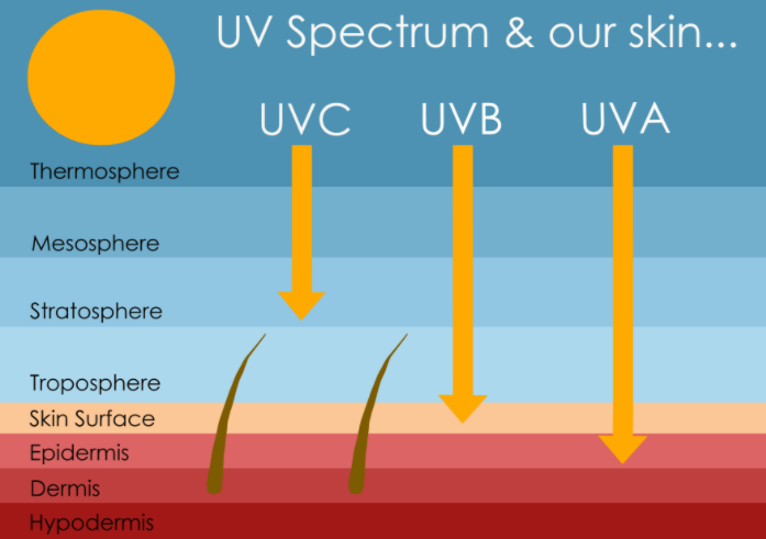
So time for another quick physics lesson. The ultraviolet (UV) radiation from the sun is broken into three parts: UVA, UVB, and UVC. Earth is enveloped by an atmosphere that protects us from radiation to a degree. UVC is unable to penetrate the atmosphere, so we’ll ignore that one. UVA can make it all the way to the ground, and UVB can make it to the ground when the sun is directly overhead. When the sun is at an angle, the UVB rays don’t make it to us. All of the harmful, DNA damaging rays come from UVB. So in other words, the mornings and afternoons are safe. A good rule of thumb to live by is that if your shadow is as tall as you, you don’t need sunscreen. If your shadow is small like it is at noon, use protection.
References:
Chakraborti, C. (2011). Vitamin d as a promising anticancer agent. Indian Journal of Pharmacology, 43(2), 113. doi:10.4103/0253-7613.77335
Duranton, F., Kramer, A., Szwarc, I., Bieber, B., Gayrard, N., Jover, B., . . . Argilés, À. (2018). Geographical variations in blood pressure level and seasonality in hemodialysis patients. Hypertension, 71(2), 289-296. doi:10.1161/hypertensionaha.117.10274
Paredes, S., Cantillo, S., Candido, K. D., & Knezevic, N. N. (2019). An Association of SEROTONIN with PAIN disorders and Its MODULATION BY ESTROGENS. International Journal of Molecular Sciences, 20(22), 5729. doi:10.3390/ijms20225729
Sintzel, M. B., Rametta, M., & Reder, A. T. (2017). Vitamin d and multiple sclerosis: A comprehensive review. Neurology and Therapy, 7(1), 59-85. doi:10.1007/s40120-017-0086-4
Other Resources:

Heart Rate Variability (HRV) and Wearable Technology
For those that like to track their health by using data and wearable technology, heart rate variability (HRV) has emerged as a new marker for resilience. HRV is the variation in the time interval between heartbeats. A low HRV has been associated with many pathologies, whereas a high HRV is associated with a healthy response to stress, and overall wellness.

For those that like to track their health by using data and wearable technology, heart rate variability (HRV) has emerged as a new marker for resilience. HRV is the variation in the time interval between heartbeats. A low HRV has been associated with many pathologies, whereas a high HRV is associated with a healthy response to stress, and overall wellness. Let’s dig into the association:
The autonomic nervous system is an unconscious system that regulates bodily functions such as heart rate, digestion, respiratory rate and several others. It controls the fight or flight response, and is divided into the sympathetic and parasympathetic systems. When the body is in a sympathetic response state, it does all the things you would want to fight or flight. Heart rate increases, the gastrointestinal system is inhibited, the pupils dilate, muscle tone increases, adrenaline is secreted. The parasympathetic nervous system’s role is to turn off that response. The muscles relax, the eyes constrict, and gastrointestinal activity resumes. The parasympathetic nervous system is controlled primarily by the vagus nerve.
Your body has a system called homeostasis, which is basically a balancing system. These systems are delicate, and many pathologies stem from an imbalance of some sort. Too much of thyroid hormone and you have Grave’s disease (hyperthyroidism), too little and you have Hashimoto’s disease (hypothyroidism). The body is striving for the goldilocks zone, and autonomic nervous system function is the same.
An interesting example of this is breathing. When you breathe in, your heart rate increases to oxygenate the tissues, and when you exhale, the heart rate decreases. There is even a difference between the nostrils. The left nostril is associated with the right brain (the creative side), and the right nostril with the left brain (the logical side). The left nostril stimulates the parasympathetic system, and the right, the sympathetic (Upadhyay-Dhungel & Sohal, 2013). Breathing with both nostrils creates homeostasis. So you can biohack this, and if you have to give a presentation and you’re nervous, plug your right nostril with your thumb and take several left sided breaths.
The vagus nerve stems from the brain and controls organs throughout the body. It calms organs after a fight or flight response, and the rate at which it can do this varies from person to person, and is a proxy for overall health. Vagal tone refers to the activity of the vagus nerve, and the body is always balancing between these two systems with vagal tone being dominant. Vagal tone can be measured indirectly through heart rate variability.
If we have constant stressors in life, whether it’s overtraining, poor diet, work related stress, or a poor night’s sleep, the balance of tone is interrupted. The ability to switch gears quickly, (moderated by the vagus nerve) is a sign of a resilient human. Regular exercise is thought to modify this ability to switch gears. It is important to note that these are associations, and if you have a low HRV, it does not necessarily mean you are in poor health, it’s more likely a snapshot of a system that is dealing with an imbalance of stress.
There are several HRV monitors on the market now, but I’m a fan of Morpheus. It can quickly track your HRV and give you a “recovery score”. With this information, you can adjust your workout accordingly. Bad night sleep and a poor score? Maybe today is not the day to try to get a PR on your squats. Fully recovered, go at it. That’s the idea. Monitoring HRV over time can give you insight into the stressors that personally affect you and you may just find yourself modifying your behaviors to get the most out of your training.
References:
Upadhyay-Dhungel, K., & Sohal, A. (2013). Physiology of nostril breathing exercises and its probable relation with nostril and cerebral dominance: A theoretical research on literature. Janaki Medical College Journal of Medical Science, 1(1), 38-47. doi:10.3126/jmcjms.v1i1.7885

Groin Pain - Ischiofemoral Impingement
Groin pain is a common complaint with many conditions of the hip, particularly in hockey players. It tends to go hand in hand with hip impingement (FAI). A lesser known condition that can cause it is called ischiofemoral impingement (IFI). This means there is a decrease in space between the femur and pelvis, and a small muscle of the hip socket (quadratus femoris) gets compressed.

Groin pain is a common complaint with many conditions of the hip, particularly in hockey players. It tends to go hand in hand with hip impingement (FAI). A lesser known condition that can cause it is called ischiofemoral impingement (IFI). This means there is a decrease in space between the femur and pelvis, and a small muscle of the hip socket (quadratus femoris) gets compressed.
What causes the decrease in space? Pelvic width plays a role which predisposes women to be at higher risk. Genu valgus (knock-kneed) could also contribute to IFI because the femur is in an adducted position. For the same reason, weakness or injury in the hip abductors can lead to IFI because it allows the femur to be in an adducted position. Lastly, surgery can be to blame. Look at the distance between the femur and the pelvis in the repaired leg.

Image from “Hip Impingement, beyond femoroacetabular” Bardakos, 2015.
How does one know if they have it? Usually resisted hip external rotation will be painful. But even more commonly, a position of adduction, external rotation, and extension will provoke pain, like so:
There is some symptom overlap with FAI as one theory of the etiology of FAI is a shortening of the external rotators of the hip from laxity in the front of the hip, as explained in this blog post. So in one scenario, we have a shortening of the quadratus femoris causing pain in the buttock and radiating pain in the groin and medial thigh, and in another scenario, we have a decrease in space compressing the same muscle causing the same symptoms. In both scenarios, the muscle has to be addressed to reduce symptoms. Here is a video on quadratus femoris release:
References:
Bardakos, N. V. (2015). Hip impingement: Beyond femoroacetabular. Journal of Hip Preservation Surgery, 2(3), 206-223. doi:10.1093/jhps/hnv049

Quadratus Femoris and Hip Impingement (FAI)
The quadratus femoris is a small rectangular muscle, deep in the hip socket. It is an external rotator and adductor of the hip. Although uncommon, tears and strains of this muscle can cause pain in the groin, posterior hip, and even cause radiating pain from irritation of the sciatic nerve.

Dancers frequently develop hip pain. Hip impingement is a common diagnosis which means there is pain in the front of the hip with movements like hip flexion and internal rotation. Usually a change in the shape of the bone in the hip is to blame, but is there more to it? We don’t know for sure, but it’s complicated.
The quadratus femoris is a small rectangular muscle, deep in the hip socket. It is an external rotator and adductor of the hip. Although uncommon, tears and strains of this muscle can cause pain in the groin, posterior hip, and even cause radiating pain from irritation of the sciatic nerve.
This set of symptoms overlaps with the pain that people experience with hip impingement. So what is the connection? With hip impingement, internal rotation of the hip is painful, and in this blog post, I explain one possible reason this pattern develops. As a person with hip impingement flexes the hip (think knee to chest), the hip will sway towards external rotation to avoid the painful internal rotation motion. The theory is that this leads to adaptive shortening of the quadratus femoris. The muscle becomes irritated and painful, and may even lead to what is known as ischiofemoral impingement (IFI), where the femur has abnormal contact with the pelvis (Gollwitzer et. al, 2017). It has been proposed that when this muscle becomes inflamed, it can leak onto the sciatic nerve which runs just posterior to this muscle and cause radiating pain down to the knee (Kassarjian et. al, 2011).

The yellow cord is the sciatic nerve, “Q” is quadratus femoris
The clinical presentation of hip impingement can be very complicated, and subsets of issues tend to come along with this diagnosis. It is difficult to determine if hip impingement may cause a shortening of the quadratus femoris, or the other way around. Regardless, lengthening this muscle and restoring range of motion of the hip, and strength in the hip abductors should be prioritized.
Here is an example of how to release quadratus femoris:
Here is an example of restoring hip internal rotation (make sure this is pain free):
And finally to strengthen the hip abductors, you can do sidelying leg raises. Here is an alternative exercise that is great for the hip abductors:
References:
Gollwitzer, H., Banke, I. J., Schauwecker, J., Gerdesmeyer, L., & Suren, C. (2017). How to address ischiofemoral impingement? Treatment algorithm and review of the literature. Journal of Hip Preservation Surgery, 4(4), 289-298. doi:10.1093/jhps/hnx035
Kassarjian, A., Tomas, X., Cerezal, L., Canga, A., & Llopis, E. (2011). MRI of THE Quadratus FEMORIS Muscle: Anatomic considerations AND Pathologic Lesions. American Journal of Roentgenology, 197(1), 170-174. doi:10.2214/ajr.10.5898

SI Joint Pain and Hip Impingement (FAI)
Hip impingement, or femoroacetabular impingement, is pain in the front of the hip that occurs with hip flexion and internal rotation. Sometimes, patients with FAI also get pain in the back side. The sacroiliac joint (SI) is usually the culprit.

Hip impingement, or femoroacetabular impingement, is pain in the front of the hip that occurs with hip flexion and internal rotation. Sometimes, patients with FAI also get pain in the back side. The sacroiliac joint (SI) is usually the culprit. To read more about the SI joint, check out this blog post.
Why does this so frequently happen? A principle of the body is that where there is an area of stiffness, the neighboring area tends to ‘pick up the slack’. In other words more strain is induced in the SI joint (Hammoud et. al, 2014). It’s similar to the shoulder. If you are missing internal rotation, and you try to reach behind your back, your whole shoulder girdle will tip forward. The same thing happens with the hip. Hip internal rotation is usually very painful and limited in FAI. So as the femur starts to rotate into internal rotation, the pelvis shifts away from the pain and creates torsion at the SI joint. This joint is embedded with a ton of ligaments and they are not contractile like a muscle, so they get inflamed from being stretched. If it gets bad enough, the inflammation ‘leaks’ onto the surrounding nerves and can send pain down the leg.
In a similar mechanism, stress can occur at pubic symphysis, the area in the front where the bones meet. Groin pain is strongly associated with FAI, and this may be the underlying mechanism. This is not to be confused with osteitis pubis, which is pain where the adductor inserts into the pubic tubercle. With osteitis pubis, pain will be elicited with flexion, abduction, and external rotation; the FABER position (Gomella & Mufarrij, 2017).
Another point of confusion is that posterior pain can also be from the hip capsule. When there is a cam deformity present, as the hip is flexed and internally rotated, the head of the femur is levered into the socket, placing stress at the back side of the capsule (Hammoud et. al, 2014). See below:
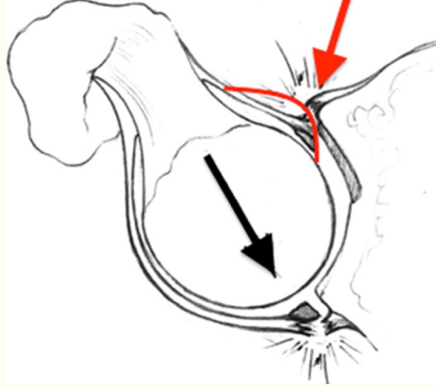
Photo taken from Hammoud et. al, 2014. The red arrow is the front and the black arrow is the back.
Seeing that lack of range of motion seems to be at the heart of the problem, restoring flexion and internal rotation will reduce strain at the SI joint. For some patients, placing a lateral distraction force helps them get into a better position. However, every person is different so this should only be done if it is pain free. Here is an example:
Think this might be you? Let’s get on a free phone call and figure it out.
References:
Gomella, P., & Mufarrij, P. (2017). Osteitis pubis: A rare cause of suprapubic Pain. Retrieved February 25, 2021, from https://www.ncbi.nlm.nih.gov/pubmed/29302238
Hammoud, S., Bedi, A., Voos, J. E., Mauro, C. S., & Kelly, B. T. (2014). The recognition and evaluation of patterns of compensatory injury in patients with Mechanical HIP PAIN. Sports Health: A Multidisciplinary Approach, 6(2), 108-118. doi:10.1177/1941738114522201
Slipped Capital Femoral Epiphysis: The Underlying Mechanism for FAI?
Slipped capital femoral epiphysis, or SCFE for short, is a growing concern. This is a condition that affects adolescents and can range from mild to severe. The hip is a ball and socket joint, with the ball being the top of the femur. Just below the ball is a growth plate, and with increased shear forces on this growth plate, the ball can slip backward and down. This is what is known as SCFE.

Slipped capital femoral epiphysis, or SCFE for short, is a growing concern. This is a condition that affects adolescents and can range from mild to severe. The hip is a ball and socket joint, with the ball being the top of the femur. Just below the ball is a growth plate, and with increased shear forces on this growth plate, the ball can slip backward and down. This is what is known as SCFE.
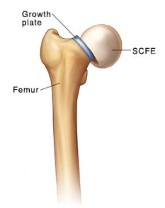
Why does this happen? Growing rates of childhood obesity are the most cited reason. It can also occur due to trauma, hormonal changes, or from chronic inflammation. It affects boys more than girls. Patients will typically present with decreased range of motion in flexion, abduction, and internal rotation. They will also usually limp and complain of pain that radiates into the knee. This is important to identify because if it is unstable and not surgically addressed, it can lead to necrosis of the bone.
There are various studies that show that as obesity rates increase, so does the rate of SCFE. For the more severe cases, the patient is surgically repaired. For more stable and less symptomatic SCFE, the patient is given crutches to allow for healing. The problem is, because the symptoms can radiate to the knee, the patient is often misdiagnosed and the SCFE progresses leading to permanent damage and bony changes. Additionally, many cases are likely not brought to attention because obese children are less active so symptoms are not elicited.
Femoral Acetabular Impingement has also garnered a lot of attention in the past decade and surgical cases are on the rise. When you look at the cam deformity, it looks a lot like an older version of SCFE.
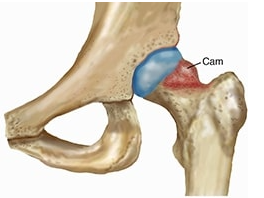
Both feature a bony protrusion on the neck of the femur. Clinically, they present the same; pain in the front of the hip with hip flexion and/or internal rotation. In 2017, Jones et. al found that increasing severity of slip deformity was a predictor of impingement and hip flexion loss, with concurrent damage to the anterosuperior aspect of the acetabulum.
It is important for clinicians that see adolescent patients to recognize this pattern to catch it before it progresses. I believe that is also incumbent upon physical therapists to have a conversation with patients and family members about dietary choices as obesity and chronic inflammation both stress the growth plate, and can be mitigated with avoidance of obesogenic and inflammatory foods.
Reference:
Jones, C. E., Cooper, A. P., Doucette, J., Buchan, L. L., Wilson, D. R., Mulpuri, K., & D’Entremont, A. G. (2017). Relationships between severity of deformity and impingement in slipped capital femoral epiphysis. Journal of Pediatric Orthopaedics, 37(4), 272-278. doi:10.1097/bpo.0000000000000641

Pain in the Front of the Hip: Subspine Impingement
Pain in the front of the hip when squatting is a common complaint, and is often diagnosed as a hip flexor problem, or hip impingement. A lesser known pathology is called subspine impingement (SSI), or anterior inferior iliac spine (AIIS) impingement. The AIIS is the bottom bony bump you can feel in the front of your pelvis and serves as a muscle attachment point. In some cases, the neck of the femur abuts against this point causing pain.

Pain in the front of the hip when squatting is a common complaint, and is often diagnosed as a hip flexor problem, or hip impingement. A lesser known pathology is called subspine impingement (SSI), or anterior inferior iliac spine (AIIS) impingement. The AIIS is the bottom bony bump you can feel in the front of your pelvis and serves as a muscle attachment point. In some cases, the neck of the femur abuts against this point causing pain.
This is slightly different from femoroacetabular impingement (FAI) in that FAI is intra-articular, meaning it occurs inside the capsule, and SSI is extra-articular. It has been described as an avulsion of the rectus femoris and its attachment to AIIS. An avulsion is when the bone pulls at the attachment site causing inflammation. It is common for patients with FAI to also have SSI (Hetsroni et. al, 2012).
Pain will typically be provoked with both active and passive hip flexion, and the patient will tend to move the hip into external rotation as the hip flexes to avoid pain. Straight leg raises will usually elicit pain, and in the early stages of rehab, a hip spica wrap may be helpful. Patients will also usually have pain with hip internal rotation and groin pain (Bech & Havercamp, 2018).
Another source of complication can be a little muscle called iliocapsularis. It lies over the front of the hip capsule and its function is unknown, but it has been theorized that its role is as a hip stabilizer. Some have described it as pulling the capsule in a superior and medial direction, and may prevent pinching in the front of the hip. Weakness in this muscle may allow for capsular pinching. One of the attachment points is the AIIS and may contribute to osteophyte (bone spur) formation, causing SSI (Sato et. al, 2016).
If you want to assess it, here is how it’s done:
References:
Bech, N. H., & Haverkamp, D. (2018). Impingement around the hip: Beyond cam and pincer. EFORT Open Reviews, 3(2), 30-38. doi:10.1302/2058-5241.3.160068
Hetsroni I, Larson CM, Dela Torre K, et al. Anterior inferior iliac spine deformity as an extra-articular source for hip impingement: a series of 10 patients treated with arthroscopic decompression. Arthroscopy. 2012; 28:1644–53.
Nabhan, D. C., Moreau, W. J., McNamara, S. C., Briggs, K. K., & Philippon, M. J. (2016). Subspine hip impingement. Current Sports Medicine Reports, 15(5), 315-319. doi:10.1249/jsr.0000000000000291
Sato T, Sato N, Sato K (2016) Review of the Iliocapsularis Muscle and its Clinical Relevance. Anat Physiol 6:237. doi: 10.4172/2161-0940.1000237

Hip Impingement and Capsular Laxity
Hip impingement has been gaining a lot of attention lately. It can be a difficult diagnosis to manage as a physical therapist, depending on severity, and can be incredibly frustrating for the patient. As more evidence comes in, we are getting a clearer picture of what exactly is the cause, and it certainly looks multifactorial. One suggested mechanism is laxity of the anterior part of the hip capsule.

Hip impingement has been gaining a lot of attention lately. It can be a difficult diagnosis to manage as a physical therapist, depending on severity, and can be incredibly frustrating for the patient. As more evidence comes in, we are getting a clearer picture of what exactly is the cause, and it certainly looks multifactorial. One suggested mechanism is laxity of the anterior part of the hip capsule.
So first, let’s understand what a capsule is. All joints have capsules around them, and they are often likened to a leathery sack. They are a blend of ligaments, connective tissue, and tendons; so they are not a unique structure. They provide stability for the joint. Depending on the position of the joint, certain parts of the capsule are stressed. With rotational movements of the joint, the anterior capsule is stressed and becomes taught. The biggest ligament in the body is the iliofemoral ligament, or Y-ligament and is in the front of the hip. It has a major role in stability. In standing, it keeps the femoral head pressed into the acetabulum. It also resists hip external rotation. Activities like gymnastics and ballet place a lot of external rotational strain on this ligament, especially in positions like the splits or turnout in ballet. Ligaments are not contractile like a muscle or tendon. Once they are stretched, they don’t go back to their original shape. They become lax.
Stability in the hip is created by several factors. There is a negative pressure inside the capsule that adds a layer of stability. In ball and sockets joints, the socket is lined with a fibrocartilage called the labrum. It deepens the socket and adds more stability. Also the muscles, tendons, and ligaments add stability. Labral tears are highly associated with hip impingement. Interestingly, the majority of labral tears in North American are in the anterosuperior part of the labrum and is associated with twisting/pivoting motions. In Asian populations, the majority of tears are posterior and are associated with hyperflexion from squatting motions (Mason, 2001). So it just may be a matter of repetition that creates laxity and leads to tearing.
So if we have a situation where we’ve created laxity in a ligament/capsule, and have a deficient labrum from tears or fraying, we have two factors that can add up to an instability that worsen each other. It’s like the wheels coming off of a bus. On top of that, there is such a focus on stretching in ballet and gymnastics which probably make a bad situation worse.
Additionally, there is what I call a “connective tissue profile”. This simply means we are all built different and there is a spectrum of elasticity to our connective tissues on an individual level. There are certainly disorders like Ehlers Danlos syndrome which is a more severe laxity, but it doesn’t have to be a syndrome for you to have more laxity than the next person. Hormones can change a ligaments integrity as well and is why there is a lot of back pain with pregnancy. Here is the other confounding factor; choice in sport. Stronger people tend to lift weights, and looser people tend to go into yoga. What I’m saying is that someone who has natural flexibility will excel in gymnastics and be drawn to that sport.
So what do we do about all of this? First, assess how lax your hip is compared to the asymptomatic one. You can do it like this:
Then aim to stabilize the joint. Remember that muscles also create stability in the joint. So you can overcompensate in a sense by strengthening the muscles surrounding the joint. I would suggest starting with the deepest ones. There is a group of muscles that are analogous to the rotator cuff of the shoulder. They do hip internal rotation and external rotation. There are 13 of them:
External rotators:
Piriformis
Gemellus Superior
Gemellus Inferior
Obturator Internus
Obturator Externus
Quadratus Femoris
Internal rotators:
Tensor fascia latae
Gluteus minimus
Gluteus medius
Adductor longus
Adductor brevis
Adductor magnus
Pectineus
It can get confusing because some of the muscles change their function based on the amount of hip flexion/extension. So the easiest thing to do is hip rotation (internal and external) in multiple planes to create maximal stability.
Here is a drill I like to do for this:
References:
Acetabular labral tears in the athlete - researchgate. (n.d.). Retrieved February 8, 2021, from https://www.researchgate.net/publication/11676689_Acetabular_labral_tears_in_the_athlete
Martin RL;Enseki KR;Draovitch P;Trapuzzano T;Philippon MJ;. (n.d.). Acetabular labral tears of the hip: Examination and diagnostic challenges. Retrieved February 08, 2021, from https://pubmed.ncbi.nlm.nih.gov/16881467/

12 Common Nutritional Deficiencies in Vegetarians
The nutrition world can be confusing and there seems to be opposite recommendations for just about any food out there. And it may be for good reason; there is not one diet approach that is perfect for everyone. With that being said, it is important to understand the pros and cons of diet styles. Most diets will be deficient in some areas which can range from minor symptoms, to more dangerous consequences.

The nutrition world can be confusing and there seems to be opposite recommendations for just about any food out there. And it may be for good reason; there is not one diet approach that is perfect for everyone. With that being said, it is important to understand the pros and cons of diet styles. Most diets will be deficient in some areas which can range from minor symptoms, to more dangerous consequences. For the purpose of brevity, this article will list the more dangerous problems that can occur because many of the milder symptoms overlap and include nausea, fatigue, weakness, confusion, etc. It is important to note that some of these nutrients can be toxic in excess, including the fat soluble vitamins, A and D.
Vitamin A: Also known as Retinol, it is important for vision. It is found mostly in animal products such as liver, eggs, dairy and oily fish, so it can be difficult for vegetarians to get a good source of vitamin A without supplementation. Deficiency can lead to eye disorders.
Vitamin D: Is involved in absorption of calcium and phosphorus to support strong bones and teeth. It also has a role in breaking down the membrane around viruses. Deficiency can lead to rickets and osteoporosis. Vitamin D is found in cod liver oil, egg yolks and red meat. Vitamin D can be produced from sunlight so it is likely more of an issue for people living in colder climates.
Leucine: Is a branched chain amino acid (BCAA) and is involved in protein synthesis and energy production. Deficiency can lead to a loss of muscle mass and hair loss. It is found mostly in meat, fish, eggs, and dairy but is also in legumes and pumpkin seeds.
Glutamine: is another amino acid and is a precursor to arginine. It is involved in protein synthesis and immune function.It is also involved in wound healing, reducing cortisol levels, and promotes gastrointestinal health. Strenuous exercise can reduce glutamine levels and lead to suppressed immune function. It is found in almost every food that is protein based.
B12: Boosts energy, helps form blood cells, and prevents brain atrophy. Dietary sources of vitamin B12 occur primarily in animal products such as lamb, beef, herring, mackerel, pork liver, oysters, poultry, clams, and eggs. To get enough B12, vegetarians have to supplement or ingest food that has been fortified with B12 such as tofu. Deficiency can lead to anemia.
Creatine: rapidly replaces ATP in fast twitch muscle fibers, improving power output. It also reduces muscle cramps. Beef is high in creatine, however when cooked, it transforms into creatinine, a waste product of creatine. Deficiency can lead to CDS (creatine deficiency syndrome) and cause developmental delays, intellectual disability, seizures, and movement disorders.
Taurine: is an amino acid that plays a role in brain tissue and nervous system functioning. It is also involved in blood pressure regulation and eye health. Deficiency can lead to weakening of the heart muscles, retinal degeneration and eventually blindness, and fetal abnormalities.
Carnosine: is synthesized from beta-alanine and L-histidine. Carnosine is involved in antioxidant activity, improves copper uptake, and reduces the fatigue caused by muscle acidosis. It is found in muscle tissue so animal products are high in carnosine. Deficiency can cause developmental delays, nerve tissue degradation, and tremors.
DHA (Docosahexaenoic Acid): is an Omega-3 fatty acid and competes with arachidonic acid, and is therefore anti-inflammatory. They also thin the blood, reduce clotting, and may raise HDL. Many children and adults with ADHD have a deficiency in DHA. Emerging evidence has linked behavior disorders with chronic inflammation of the brain. DHA is found in fish oils and grass fed beef.
Heme Iron: is crucial in delivering oxygen to our tissues of the body. Although you can get iron from plants, it is non-heme iron and less absorbable in the body. Iron deficiency can lead to anemia which can cause fatigue, headaches, restless legs, and oddly enough, the desire to chew ice.
Calcium: is important in bone health and controlling blood pressure. It is important to have zinc and vitamin D in the diet to absorb and use calcium, and these tend to be deficient on a plant-based diet. Calcium deficiency can cause osteoporosis, tingling, cramps, and spasms.
Zinc: is important in growth and repair of muscle. It also has a role in the immune system and prevents RNA replication in viruses. Deficiency can lead to hair loss, and lack of smell and taste.
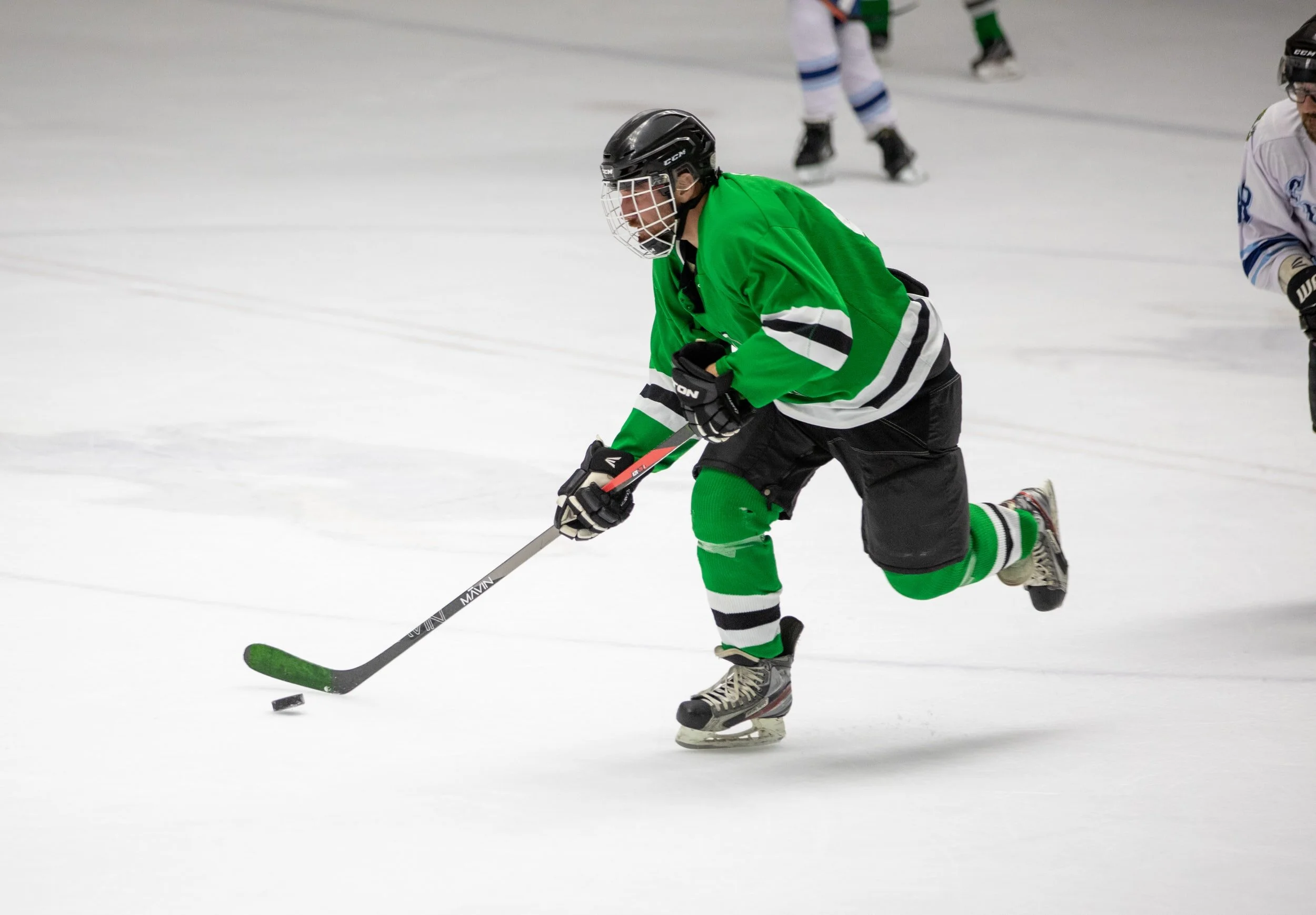
Hockey: 9 Exercises To Add Velocity To Your Wrist Shot
Want to improve your velocity? Let’s be honest, probably every muscle in the body is active when shooting a puck, but by focusing on the prime movers, you can easily gain explosive speed.

Want to improve your velocity? Let’s be honest, probably every muscle in the body is active when shooting a puck, but by focusing on the prime movers, you can easily gain explosive speed. Let’s break down the movements and muscles involved from the ice up, from a right handed shooter’s perspective.
1. The right hip has to abduct and externally rotate which is mostly gluteus medius, minimus, and piriformis. Hip airplanes are a great way to target these muscles:
2. The spine has to rotate which involves the spinal rotators and obliques. Single leg chops hit these muscles and get the glutes involved also:
3. The right shoulder needs to adduct and internally rotate. For this, try some flies from low to high:
4. The left shoulder needs to externally rotate. Classic shoulder external rotation will work but from an angled position:
5. The left shoulder also adducts. You can use shoulder adduction for this:
6. The right forearm pronates forcefully. To target pronator teres and quadratus, try this:
7. The left forearm has to supinate which involves the bicep and the supinator. For the bicep, curls will do the job but add supination:
8.Here is a great way to target the supinator muscle:
9. Grip strength and forearm strength are always going to help for a quick shot. Here is an oldie but goodie:
A couple of tips:
Always train bilaterally, so do all these on both sides. Sports like hockey, golf, and baseball that are highly rotational in one direction will lead to imbalances later if not trained bilaterally.
Sports are all about rate of force production. That means quick movement. So I’d recommend performing the concentric movement (positive phase) quickly, then perform the eccentric movement (negative phase) slowly.
You can use cables for a lot of these motions but there is an advantage that elastic bands have. When it comes to range of motion, most muscles are weakest at the end of a range of motion. An elastic band has more tension towards the end range of motion so it specifically targets the end range.
Want to add more specificity to your programming? Check out our Train Smarter Program:

Hip Impingement and Paralabral Cysts
A common finding in patients with hip impingement, or FAI, is a labral tear. Where there is a labral tear, there is frequently a paralabral cyst. In fact, cysts are seen on MRIs between 50-70% of patients with labral tears, but only 5% in the normal population

A common finding in patients with hip impingement, or FAI, is a labral tear. Where there is a labral tear, there is frequently a paralabral cyst. In fact, cysts are seen on MRIs between 50-70% of patients with labral tears, but only 5% in the normal population (Shin et al., 2017). The association is so strong that the presence of a paralabral cyst can indirectly be used as a sign of a labral tear (Magee & Hinson, 2000). Many times these cysts do not cause pain, but for an unlucky few, it can press on neurovascular bundles and cause pain and paresthesia into the leg. It is not well understood, but it appears to be all about location.
The cause of these cysts is unknown, but is commonly seen where there is injury. One line of thought is that a torn labrum allows for synovial fluid to “leak” causing a collection and a fluid filled sack.
A symptomatic cyst will have the same pattern of pain as a labral tear. Pain with flexion, adduction, and internal rotation. Because of this position, hockey players are susceptible as well as anyone who is in a sustained flexed position.
There is limited studies on paralabral cysts, and most are in the form of case reports. One case report described a patient that had a labral tear and a cyst about half the size of a penny on the back side of the joint. The patient was experiencing pain, and tingling down the leg, a.k.a. sciatica. All other pathologies were ruled out, and the patient had the cyst removed and the labrum repaired. It was considered a success and the patient was pain free following the surgery (Salunke & Panchal, 2014).
The good news is that most cysts of the hip occur towards the front of the hip and only 17% occur on the back side, near the sciatic nerve (El-Feky & Batta, 2021). So most of the time, they do not cause problems, but for the unlucky few, surgery or aspiration may be recommended.
References:
Batta, N., & El-Feky, M. (n.d.). Paralabral cyst of the hip: Radiology Reference Article. Retrieved January 19, 2021, from https://radiopaedia.org/articles/paralabral-cyst-of-the-hip-1
Magee, T., & Hinson, G. (2000). Association of Paralabral Cysts with Acetabular Disorders. American Journal of Roentgenology, 174(5), 1381-1384. doi:10.2214/ajr.174.5.1741381
Salunke, A., & Panchal. (2014, July 21). A Paralabral Cyst of the Hip Joint Causing Sciatica: Case Report and Review of Literature. Retrieved January 19, 2021, from http://europepmc.org/articles/PMC4418116
Shin, K., Park, S., & Lee, W. (2017). Paralabral Cyst of the Hip Compressing Common Femoral Vein Treated with Sono-guided Cyst Aspiration Followed by Arthroscopic Labral Debridement: A Case Report. Hip & Pelvis, 29(3), 194. doi:10.5371/hp.2017.29.3.194

Sugar and Arthritis
If you have knee arthritis, I’m sure you’ve been told that you should lose weight. For decades, we’ve assumed that arthritis is caused from “wear and tear” and is just caused by repetitive mechanical loading. So if you’re overweight, the joints take on more compression, eroding cartilage faster. But are we sure that is all that is going on?

If you have knee arthritis, I’m sure you’ve been told that you should lose weight. For decades, we’ve assumed that arthritis is caused from “wear and tear” and is just caused by repetitive mechanical loading. So if you’re overweight, the joints take on more compression, eroding cartilage faster. But are we sure that is all that is going on?
If mechanical compression causes arthritis, then how do we explain the piles of evidence that running is good for your knees, not bad. When it comes to research, there are varying degrees of evidence. Randomized controlled trials (RCTs) are pretty top notch, but even better than that is a systematic review. This means that all of the RCTs on a certain topic are summarized. A 2011 systematic review (Urquhart et al.) on physical activity and knee joint health found that “physical activity is beneficial, rather than detrimental, to joint health”. They go on to say that physical activity is also associated with an increase in cartilage volume (yes, increase) and a decrease in cartilage defects.
We do see that when someone loses just 10% of their body mass, they experienced significant pain resolution or reduction, reduced inflammation, and improved function (Messier et al., 2013). We have assumed that it just decreased mechanical compression, but are we sure?
We also know that diabetics, who tend to have high glucose concentrations, are at high risk for early onset arthritis. In 2016, (Courties & Sellam) published an article that surmised that low grade inflammation and the induction of oxidative stress and cytokines contribute to joint related pain.
When sugar is in close proximity to a protein, it attaches to it. This is a process known as glycation. This is what is being measured in a HbA1c test. Once the protein is glycated, it changes to advanced glycation end products (AGEs). These AGEs are able to crosslink proteins and cause collagen stiffness. This leads to tissue remodeling and degradation (Degroot et al., 2014).
The good news is that this goes both ways. A low carb, high protein diet can improve tissue remodelling of the extracellular matrix and improve tissue quality. An RCT published in 2019 (Strath et al.), found that low carb diets are more effective than low fat diets in reducing pain intensity from knee arthritis.
I intentionally chose the picture for this article to include table sugar and strawberries. This is because when I say sugar, I really mean carbohydrates. That is glucose, sucrose, galactose, fructose, etc. Now I don’t mean to vilify carbs, but it is well known that the average Americans consumes an over abundance of carbs, partly because we have been told for decades to “carb up”. Still to this day, dietary guidelines are still heavy on the carb portion. Some of the current ideas behind new diets like the carnivore diet, is to eat like our ancestors. If you think about it, our ancestors didn’t have fruits and vegetables around all the time, and their diet was mostly meat-centric. Not to say that fruits and vegetables are bad, rather we probably take in more than our ancestors did.
So the next time your achy knee acts up, instead of rushing out to get an injection or swallowing some anti-inflammatories, try cutting back on the carbs for a week. It may be all that you need.
References:
Courties, A., & Sellam, J. (2016). Osteoarthritis and type 2 diabetes mellitus: What are the links? Diabetes Research and Clinical Practice, 122, 198-206. doi:10.1016/j.diabres.2016.10.021
Degroot, J., Verzijl, N., Wijk, M. J., Jacobs, K. M., El, B. V., Roermund, P. M., . . . Lafeber, F. P. (2004). Accumulation of advanced glycation end products as a molecular mechanism for aging as a risk factor in osteoarthritis. Arthritis & Rheumatism, 50(4), 1207-1215. doi:10.1002/art.20170
Messier, S. P., Mihalko, S. L., Legault, C., Miller, G. D., Nicklas, B. J., Devita, P., . . . Loeser, R. F. (2013). Effects of Intensive Diet and Exercise on Knee Joint Loads, Inflammation, and Clinical Outcomes Among Overweight and Obese Adults With Knee Osteoarthritis. Jama, 310(12), 1263. doi:10.1001/jama.2013.277669
Strath, L. J., Jones, C. D., George, A. P., Lukens, S. L., Morrison, S. A., Soleymani, T., . . . Sorge, R. E. (2019). The Effect of Low-Carbohydrate and Low-Fat Diets on Pain in Individuals with Knee Osteoarthritis. Pain Medicine, 21(1), 150-160. doi:10.1093/pm/pnz022
Urquhart, D. M., Tobing, J. F., Hanna, F. S., Berry, P., Wluka, A. E., Ding, C., & Cicuttini, F. M. (2011). What Is the Effect of Physical Activity on the Knee Joint? A Systematic Review. Medicine & Science in Sports & Exercise, 43(3), 432-442. doi:10.1249/mss.0b013e3181ef5bf8

Weight Loss and Iron Deficiency
Do you crave ice? Do you have restless leg syndrome? Or maybe you have chronic fatigue and look pale. Do you get short of breath? Do you bruise easily? These are all signs of iron deficiency. Did you know a deficiency in iron can inhibit your ability to lose weight? Let’s dig into it.
Do you crave ice? Do you have restless leg syndrome? Or maybe you have chronic fatigue and look pale. Do you get short of breath? Do you bruise easily? These are all signs of iron deficiency. Did you know a deficiency in iron can inhibit your ability to lose weight? Let’s dig into it.
Iron deficiency is extremely common, and it is a type of anemia. According to webmd, It affects about 20% of women, 50% of pregnant women, and 3% of men. Part of the reason that women are more affected is that women tend to eat less red meat. Another reason is blood loss during menstruation, and increased need for iron (because you are supporting another life) during pregnancy.
With the movement towards plant-based eating, the problem will certainly rise. Iron comes in two forms; heme iron (from red meats) and non-heme iron. Although you can get some iron from plants, it comes in non-heme iron form, which is less well absorbed by the body. Additionally, plants contain polyphenols, which inhibit absorption of iron. Vitamin C can enhance absorption of iron (Ems, Lucia & Huecker, 2020).
Why is iron important? Many proteins and enzymes in the body require iron to function. A vital protein involved in energy pathways of the body (electron transfer) is called cytochrome. With iron deficiency, cytochrome is unable to do its job and energy production plummets (Blayney et al., 1976). This is the reason for fatigue in iron deficiency. If you’re fatigued, it’s difficult to get your exercise in and will limit weight loss.
Another interesting point on iron is its relationship with inflammation. Iron is stored in the body by a molecule called ferritin. Bacterial replication requires iron, so when you have an infection, the body wisely locks the ferritin so that the bacteria cannot use it to replicate. Although this is a protective process and necessary, now the body cannot use iron. A similar process occurs with chronic inflammation (Ueda and Takasawa, 2018). So chronic inflammatory processes like diabetes, obesity, and auto-immune disease can lead to chronic iron deficiency. This is called functional iron deficiency.
So if you eat red meat, the darker the better. The pigment of the meat is from myoglobin, which contains heme iron, not from the color of blood. If you don’t eat red meat or are on a plant-based diet, it will be important to consult with a physician to ensure proper iron levels. If you have a chronic illness causing inflammation, the root cause of the inflammation will need to be treated first.
References:
Blayney, L., Bailey-Wood, R., Jacobs, A., Henderson, A., & Muir, J. (1976). The effects of iron deficiency on the respiratory function and cytochrome content of rat heart mitochondria. Circulation Research, 39(5), 744-748. doi:10.1161/01.res.39.5.744
MR;, E. (n.d.). Biochemistry, Iron Absorption. Retrieved January 06, 2021, from https://pubmed.ncbi.nlm.nih.gov/28846259/
Ueda, N., & Takasawa, K. (2018). Impact of Inflammation on Ferritin, Hepcidin and the Management of Iron Deficiency Anemia in Chronic Kidney Disease. Nutrients, 10(9), 1173. doi:10.3390/nu10091173

Pain on the Top of the Foot: Extensor Digitorum
A frequent complaint from runners is pain on the top of the foot. Although things like stress fractures can occur, it’s not common unless there is a traumatic event. More often than not, it’s a problem with the extensor mechanism of the toes. Either the tendons get inflamed, or the sheath they slide in and out of gets inflamed. The muscle in question in the extensor digitorum longus.

A frequent complaint from runners is pain on the top of the foot. Although things like stress fractures can occur, it’s not common unless there is a traumatic event. More often than not, it’s a problem with the extensor mechanism of the toes. Either the tendons get inflamed, or the sheath they slide in and out of gets inflamed. The muscle in question in the extensor digitorum longus. Here is what it looks like:
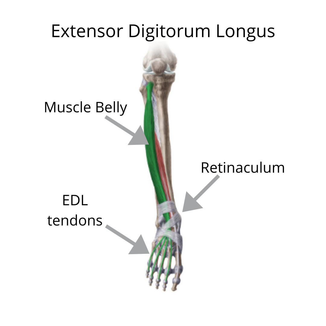
You’ll notice that the tendons insert on all of the toes except the big toe. There is also a retinaculum which is a piece of connective tissue on the front of the ankle that keeps the tendons from bowstringing, and acts as a fulcrum point. The muscle acts to lift the toes up, assists with dorsiflexion and eversion of the foot.
If you are swollen and bruised, you should consult with a physician. When there is bruising, blood vessels have been interrupted, and is a sign (although not always) of tissue damage. This could be rupture of a muscle or ligament, or a stress fracture. If it’s just swollen and not bruised, it’s more likely that you have an overuse injury such as a tendonitis or tenosynovitis.
So why does this happen? This can occur when running hills, up stairs, or in the case of a friend of mine, when running the warped wall in a ninja obstacle course. All of these motions require you to forcefully lift your toes and foot upward so you don’t trip. There are, of course, contributing factors that can lead to any overuse injury, and with ankle injuries, lack of dorsiflexion is usually an underlying culprit.
With this particular situation, lack of dorsiflexion will lead to foot eversion. In other words, your foot will turn out when you run to make up for the lack of motion required to run. This puts the extensor digitorum into a shortened position which is a great way to strain it. Also, if you are restricted in dorsiflexion, the muscles in the front of the foot, including extensor digitorum, have to fight against the tissue resistance, also leading to strain. It’s like running with a hand brake on. So the long term fix will be to restore your ankle dorsiflexion and work on your landing mechanics so that you run and land with your foot straight.
But if you are dealing with this, and it can be exquisitely painful, you will first want to get out of pain and feeling normal. Here is where we use the D2R2 model:
Step 1: (D1) Desensitize. This can be done with massage, cupping, scraping, etc. Anything to reduce pain levels
Step 2: (D2) Decongest. Get the static fluid out of the area. Intermittent compression works great with this step. We recommend voodoo flossing like so:
You can also do the same thing with the wrap going over the top of the foot to address the tendons. If you are still swollen, use the effects of gravity. Lay on your back with your legs up on a wall. Pump your ankles 30 times. Then contract your quads 30 times, then contract your glutes 30 times, then take 10-20 deep breaths. With each inhale, make sure your belly rises, and then descends on exhale. This is a great way to get movement through your lymphatic system which is your drainage system.
Step 3: (R1) Reperfuse. Get new blood into the area. You can do this with pain free movement.
Step 4: (R2) Restore. Normalize your range of motion and strength to the area.
Here is a video of steps 3 and 4:
Once everything is back to normal, start working on getting full ankle dorsiflexion back like so:
Want to work on your ankles at home? Check out our ankle program.

Quad Tightness and Knee Pain
If you have pain in the knee with squatting or running, check out your quadriceps. There are several reasons for knee pain but an easy one to address is quad tightness. If the quad is restricted, it can cause the patella (knee cap) to compress into the femur. This is problematic because the underside of the patella is lined with cartilage and over time you can erode the cartilage, leading to arthritis.

If you have pain in the knee with squatting or running, check out your quadriceps. There are several reasons for knee pain but an easy one to address is quad tightness. If the quad is restricted, it can cause the patella (knee cap) to compress into the femur. This is problematic because the underside of the patella is lined with cartilage and over time you can erode the cartilage, leading to arthritis.
First, determine exactly where the pain is. If it’s in the joint line just below the patella, it could be meniscus and should be checked out. If it’s further up, like the area where the quad meets the patella, it could be muscular. One quick test is to lay on your back, grab your ankle with your hands, and bend your knee to your chest. You should be able to bend the knee enough for your calf to touch your hamstring. Or, when laying on your stomach, you should be able to get your foot to your butt (using a strap) without discomfort. If you can’t get there, or it causes pain, or your butt lifts up, these are all signs of quad tightness.

This would be an example of quad tightness because this hips lift off of the table. The reason this happens is that part of the quad attaches to the front of the pelvis. The pelvis tilts anteriorly as a way to give slack to the muscle. This also demonstrates the relationship of quad tightness and back pain. If the lumbar spine is sensitive to extension and the quad is tight, the person will experience back pain with deep knee bending.
Here is something you can try to increase mobility:
When I find a stiff quad, I almost always find some stiff heel cords and limited ankle range of motion. This pattern can significantly restrict your ability to squat or run. If you want to improve ankle mobility, check out this program we’ve put together.
We also have a free Ebook on running, see below:
Fill this form out to receive the Ebook.

Tibialis Posterior Tendonitis
Most problems of the foot and ankle can be drawn back to lack of ankle dorsiflexion, and tibialis posterior tendonitis is no exception. It can be excruciating, and mistaken for other pathologies, even knee pain or meniscus tears.

Most problems of the foot and ankle can be drawn back to lack of ankle dorsiflexion, and tibialis posterior tendonitis is no exception. It can be excruciating, and mistaken for other pathologies, even knee pain or meniscus tears. The origin of the muscle is just below the knee, runs down the shin medially (inside), takes a 90 degree turn behind the ankle, and inserts into the arch. It is an ankle inverter and plantarflexor, but more importantly, it helps form the arch.
If you are missing ankle dorsiflexion, your foot will turn outward when you walk. As you are toeing off, your heel whips back to make up for the outward position. This motion biases overuse of posterior tibialis.
Furthermore, if you are flatfooted, the inside ankle puts pressure on the tendon like a bowstring, causing more irritation. Now, this is a bit of a chicken or the egg scenario. Because of the role this muscle in creating an arch, you can make the case that an inefficient tibialis posterior causes a flat foot. Or, that a flat foot creates overuse of tibialis posterior and puts pressure on it. Either way, the treatment is the same. Reduce pain first, so you can function. Isometrics are good at cooling down tendonitis. Try this simple exercise:
Then you want to restore dorsiflexion. Try these two exercises:
Also, having a flat foot is not a life sentence. You can improve your arch through strengthening tibialis posterior when the pain is gone, and by strengthening the foot intrinsic muscles. Try this out:
You can try an orthotic initially, to form an arch and reduce pressure on the muscle. However, you should aim to rebuild your arch through exercise and eventually ditch the orthotic.
Want to work on your ankles at home? Check out our ankle program:

Sleep Apnea and Alzheimer’s Disease
Most people are aware of the repercussions of sleep apnea such as increased risk of hypertension and stroke, but this is a strong association that needs to be talked about. If you chronically snore or choke in your sleep, you must get it evaluated as the emerging evidence just becomes stronger that sleep apnea is a precursor to Alzheimer’s disease.
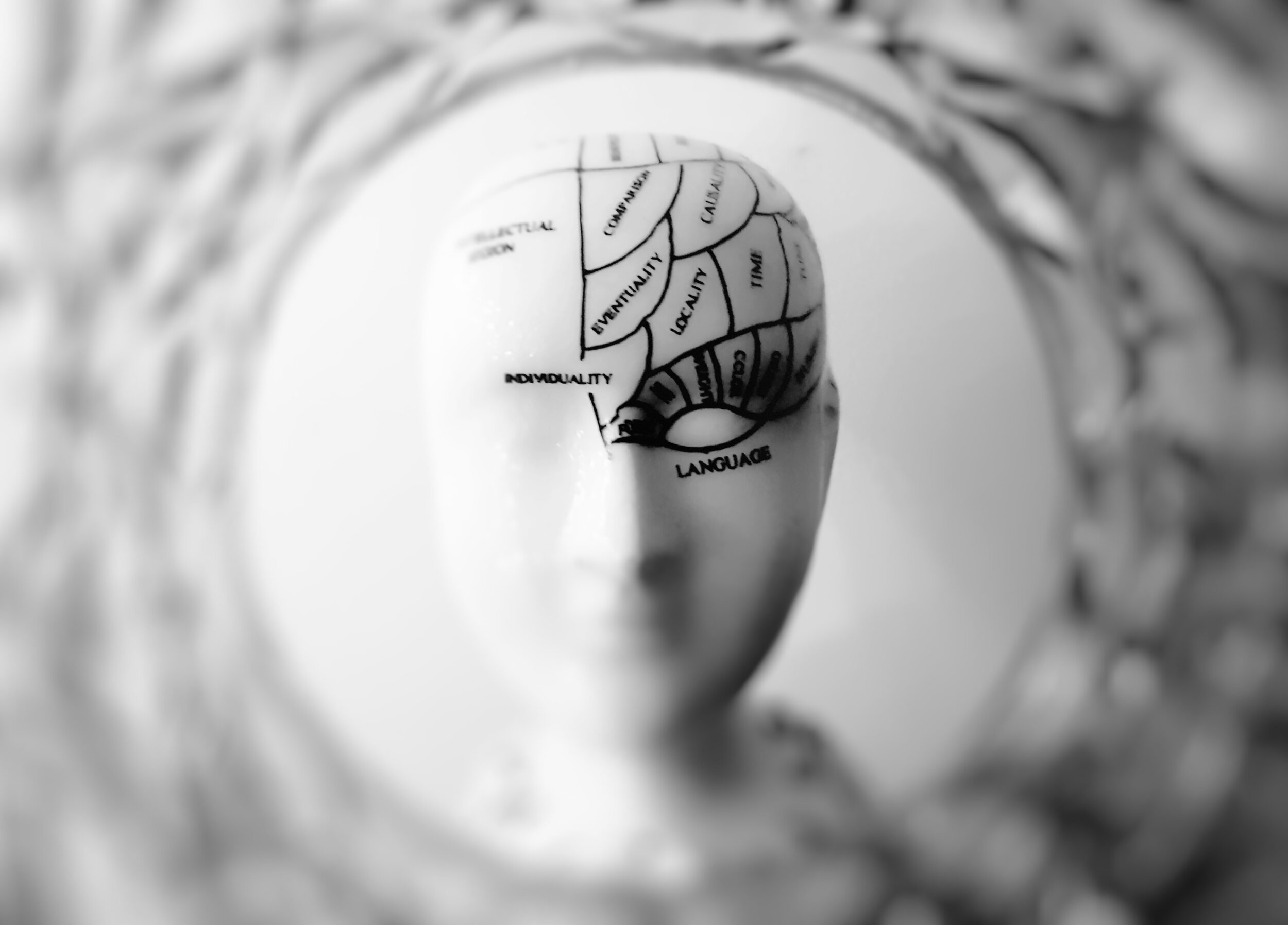
Last week we looked at the lymphatic system which has a role in filtering the blood and in immune response. The ‘sewage system’ of the body. There is an analogous system in the brain called the glymphatic system.
The glymphatic system got its name from a portmanteau of lymphatic and glial, which are nerve cells in the brain that are associated with glymph. The glymphatic system removes interstitial fluid and debris. Glymphatic activity is low during waking hours, but picks up its activity during sleep (Hauglund et al., 2020). It peaks during the deeper stages of sleep.
There is a protein, amyloid-β, that has a sticky quality. Accumulation of this protein has been associated with neurodegenerative disease such as Alzheimer’s. In fact, 25-60% of patients with Alzheimer’s disease have a sleep dysfunction (Hauglund et al., 2020). Sleep apnea is common and affects 22% of the population (Malhotra, 2009), that’s about 56 million Americans.
Most people are aware of the repercussions of sleep apnea such as increased risk of hypertension and stroke, but this is a strong association that needs to be talked about. If you chronically snore or choke in your sleep, you must get it evaluated as the emerging evidence just becomes stronger that sleep apnea is a precursor to Alzheimer’s disease.
References:
Hauglund, N. L., Pavan, C., & Nedergaard, M. (2020). Cleaning the sleeping brain – the potential restorative function of the glymphatic system. Current Opinion in Physiology, 15, 1-6. doi:10.1016/j.cophys.2019.10.020
Malhotra, A. (2009). Obstructive Sleep Apnea and Central Sleep Apnea: Epidemiology, Pathophysiology, and Risk Factors. ACCP Sleep Medicine Board Review: 4th Edition, 193-200. doi:10.1378/smbr.4th.193
Diaphragmatic Breathing and The Lymphatic System
The lymphatic system is a vital system of the body that is often overlooked. For some reason, in every textbook it is depicted as green. In actuality, it is clear or murky white from proteins and lipids. It looks a lot like the vascular system and that is no mistake. Its main function is filtration of blood plasma in the capillary beds. So where the vascular system goes, so does the lymphatic system.
The lymphatic system is a vital system of the body that is often overlooked. For some reason, in every textbook it is depicted as green. In actuality, it is clear or murky white from proteins and lipids. It looks a lot like the vascular system and that is no mistake. Its main function is filtration of blood plasma in the capillary beds. So where the vascular system goes, so does the lymphatic system. It returns 3 liters of fluid to the blood every day. In this process, cellular debris and waste is filtered out. The lymphatic system also has a role in immune response, releasing T-cells from the bone marrow to the thymus to neutralize pathogens.
The lymphatic system is one continuous system, and if something is dysfunctional in the system, you get back up in the form of swelling. Muscular contraction causes movement of the lymphatic system. That is why when you are sedentary, like on an airplane, your socks leave indentations. Because of gravity and the lack of movement, your ankles swell.
One key difference between the lymphatic system and the cardiovascular system is the heart. The heart is a pump that facilitates the circulation of blood. The lymphatic system does not have a pump. On top of that, the lymph is drained in the chest so it has to move against gravity. More specifically, the right upper quadrant drains in the upper right chest, and the rest of the body drains in the left upper chest.
There is a specialized part of the lymphatic system called the cisterna chyli. It is the most common drainage trunk of the lymphatic system and facilitates drainage for the whole body besides the upper right quadrant. It resides in the upper lumbar vertebrae. This is a good thing because as you inhale, the abdominal cavity and lungs fill, and press on the cisterna chyli and the thoracic duct, pushing excess fluid off to be drained. In other words, breathing (particularly diaphragmatic breathing) is one of the key ‘pumps’ of the lymphatic system.
Another way to facilitate lymphatic drainage is through movement. As stated above, muscular contraction pushes fluids through the system. If you’ve ever seen someone in a shoulder sling, there is usually a ball at the end of it for them to squeeze. It’s not about strengthening the grip, it more acts as a mechanical pump for a swollen shoulder. Vertical movement is particularly good, and I recommend jumping rope for lymphatic movement.
So if you just banged your elbow and it is swollen, go for a walk. Maybe lay down afterwards and work on some breathing. These simple things can have a dramatic effect on inflammation.